Publications

2024

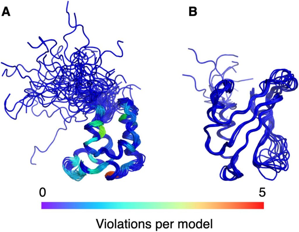
Baskaran H, Ploskon E, Tejero R, Yokochi M, Harrus D, Liang Y, …Montelione GT*, Vuister GW*, Young JY*.   Restraint validation of biomolecular structures determined by NMR in the Protein Data Bank.  Structure (Cell Press) Preprint.

Klukowski P, Damberger FF, Allain FH, Iwai H, Kadavath H, Ramelot TA, Montelione GT, Riek R, Güntert P. The 100-protein NMR spectra dataset: A resource for biomolecular NMR data analysis. Scientific Data. 2024 Jan 4;11(1):30.

Gutsche I, Montelione GT. Editorial overview: Biophysical methods: Multiple structures of proteins underpin their biological functions. Curr Opin Struct Biol. 2024 Jan 12;84:102762. doi: 10.1016/j.sbi.2023.102762. Epub ahead of print. PMID: 38217897.

2023

Kryshtafovych A, Montelione GT, Rigden DJ, Mesdaghi S, Karaca  E, Moult J. Breaking the conformational ensemble barrier: Ensemble structure modeling challenges in CASP15. PROTEINS: Structure Function Bioinformatics Online. 1-9. doi:10.1002/prot.26584. 2023.

Ramelot TA, Tejero R, Montelione GT. Representing structures of the multiple conformational states of proteins. Curr. Opin. Struct. Biol. 83, 102703. 2023.

Mazzei L, Greene-Cramer R, Bafna K, Jovanovic A, De Falco A, Acton TB, Royer CA, Ciurli S, Montelione GT. Protocol for production and purification of SARS-CoV-2 3CLpro. STAR Protocols. 2023 May 5;4(2):102326-.

Ramelot TA, Palmer J, Montelione GT, Bhardwaj G. Cell-permeable chameleonic peptides: Exploiting conformational dynamics in de novo cyclic peptide design. Current Opinion in Structural Biology. 2023 Jun 1;80:102603.

Li EH, Spaman LE, Tejero R, Huang YJ, Ramelot TA, Fraga KJ, Prestegard JH, Kennedy MA, Montelione GT. Blind assessment of monomeric AlphaFold2 protein structure models with experimental NMR data. Journal of Magnetic Resonance. 2023 May 20:107481.

Maisuradze GG, Montelione GT, Rackovsky S, Skolnick J. Protein Folding and Dynamics─ An Overview on the Occasion of Harold Scheraga’s 100th Birthday. The Journal of Physical chemistry. B. 2023 Apr 1;127(13):2879-80.

Mondal A, Swapna GV, Lopez MM, Klang L, Hao J, Ma L, Roth MJ, Montelione GT, Perez A. Structure Determination of Challenging Protein–Peptide Complexes Combining NMR Chemical Shift Data and Molecular Dynamics Simulations. ACS J. Chem Inform Modeling, 2023.. 2023 Mar 29;63(7):2058-72.

2022

Khushboo Bafna, Christopher L Cioffi, Robert M Krug, Gaetano T Montelione. Structural similarities between SARS-CoV2 3CLpro and other viral proteases suggest potential lead molecules for developing broad spectrum antivirals. Frontiers in Chemistry. 2022; 10: 948553.

Hu K, Lee W, Montelione GT, Sgourakis NG, Vögeli B. Computational approaches for interpreting experimental data and understanding protein structure, dynamics and function relationships. Frontiers in Molecular Biosciences. 2022;9.

Bhardwaj, Gaurav, O’Connor, Jacob, Rettie, Stephen, Huang, Yen-Hua, Ramelot, Theresa A., Mulligan, Vikram Khipple, Alpkilic, Gizem Gokce, Palmer, Jonathan, Bera, Asim K., Bick, Matthew J., Di Piazza, Maddalena, Li, Xinting, Hosseinzadeh, Parisa, Craven, Timothy W., Tejero, Roberto, Lauko, Anna, Choi, Ryan, Glynn, Calina, Dong, Linlin, Griffin, Robert, van Voorhis, Wesley C., Rodriguez, Jose, Stewart, Lance, Montelione, Gaetano T., Craik, David, and Baker, David. Accurate de novo design of membrane-traversing macrocycles. Cell. 2022 Sep 15;185(19):3520-32.

Keith Jeffrey Fraga, Yuanpeng J Huang, Theresa A Ramelot, GVT Swapna, Arwin Lashawn Anak Kendary, Ethan Li, Ian Korf, Gaetano T Montelione. SpecDB: A relational database for archiving biomolecular NMR spectral data. Journal of Magnetic Resonance. 2022. 107268, ISSN 1090-7807.

Yaohui Li, Rongzhen Zhang, Chi Wang, Farhad Forouhar, Oliver B Clarke, Sergey Vorobiev, Shikha Singh, Gaetano T Montelione, Thomas Szyperski, Yan Xu, John F Hunt. Oligomeric interactions maintain active-site structure in a noncooperative enzyme family. The EMBO Journal. 2022, 41: e108368.

Roberto Tejero, Yuanpeng Janet Huang, Theresa A Ramelot, Gaetano T Montelione. AlphaFold Models of Small Proteins Rival the Accuracy of Solution NMR Structures. Frontiers in Molecular Biosciences. 9, 877000. 2022.

Yuan Wang, Bochao Liu, Huimei Lu, Jingmei Liu, Peter J Romanienko, Gaetano T Montelione, Zhiyuan Shen. SETD4-mediated KU70 methylation suppresses apoptosis. Cell Reports. 2022, 39, 110794.

Daniel R Colman, Gilles Labesse, Gurla VT Swapna, Johanna Stefanakis, Gaetano T Montelione, Eric S Boyd, Catherine A Royer. Structural evolution of the ancient enzyme, dissimilatory sulfite reductase. Proteins: Structure, Function, and Bioinformatics. 2022,90:1331–1345.

2021

Anishchenko I; Pellock SJ; Chidyausiku TM; Ramelot TA,;Ovchinnikov S; Hao J; Bafna K; Norn C; Kang A; Bera AK; DiMaio F; Carter L; Chow CM; Montelione GT; Baker D. De novo protein design by deep network hallucination. Nature, 2021, 600: 547–552.

Huang YJ, Zhang N, Bersch B, Fidelis K, Inouye M, Ishida Y, Kryshtafovych A, Kobayashi N, Kuroda Y, Liu G, LiWang A, Swapna GVT, Wu N, Yamazaki T, Montelione GT. Assessment of prediction methods for protein structures determined by NMR in CASP14: Impact of AlphaFold2. Proteins: Structure, Function, and Bioinformatics. 2021 Dec;89(12):1959-76..

Aiyer S, Swapna GV, Ma LC, Liu G, Hao J, Chalmers G, Jacobs BC, Montelione GT, Roth MJ. A common binding motif in the ET domain of BRD3 forms polymorphic structural interfaces with host and viral proteins. Structure. 2021 Aug 5;29(8):886-98.

Koga N; Koga R; Liu G; Castellanos J; Montelione GT; Baker D. Role of backbone strain in de novo design of complex alpha/beta protein structures. Nature Communication. 2021, 12: 3921.

Mechanism of synergy

Bafna K; White K; Harish B; Rosales R; Ramelot TA; Acton TB; Moreno E; Kehrer T; Miorin L; Royer CA; Garcia-Sastre A; Krug RM; Montelione GT. Hepatitis C virus drugs that inhibit SARS-CoV-2 papain-like protease synergize with remdesivir to suppress viral replication in cell culture. Cell Reports. 2021, 35: 109133.

Cole CA, Daigham NS, Liu G, Montelione GT, Valafar H. REDCRAFT: A computational platform using residual dipolar coupling NMR data for determining structures of perdeuterated proteins in solution. PLoS computational biology. 2021 Feb 1;17(2):e1008060.

Mehla J, Liechti G, Morgenstein RM, Caufield JH, Hosseinnia A, Gagarinova A, Phanse S, Goodacre N, Brockett M, Sakhawalkar N, Babu M, Xiao Rong, Montelione GT, Vorobiev S, den Blaauwen T, Hunt JF, Uetz P. ZapG (YhcB/DUF1043), a novel cell division protein in gamma-proteobacteria linking the Z-ring to septal peptidoglycan synthesis. Journal of Biological Chemistry. 2021 Jan 1;296.

2020

Maisuradze GG, Montelione GT, Rackovsky S, Skolnick J. Tribute to Harold A. Scheraga. The Journal of Physical Chemistry B. 2020 Oct 23;124(46):10301-2.

Bafna K; Krug RM; Montelione GT. Structural similarity of SARS-CoV2 Mpro and HCV NS3/4A proteases suggests new approaches for identifying existing drugs useful as COVID-19 therapeutics. ChemRxiv. 2020. (Preprint Version)

Chen, G., Ma, L.-C., Wang, S., Woltz, R.L., Grasso, E.M., Montelione, G.T., Krug, R.M. A double-stranded RNA platform is required for the interaction between a host restriction factor and the NS1 protein of influenza A virus.  Nucleic Acids Research. 48: 304-315, 2020.

Wang X, Jing X, Deng Y, Nie Y, Xu F, Xu Y, Zhao YL, Hunt JF, Montelione GT, Szyperski T. Evolutionary coupling saturation mutagenesis: Coevolution‐guided identification of distant sites influencing Bacillus naganoensis pullulanase activity. FEBS letters. 2020 Mar;594(5):799-812.

2019

Berman HM, Adams PD, Bonvin AA, Burley SK, Carragher B, Chiu W, DiMaio F, Ferrin TE, Gabanyi MJ, Goddard TD, Griffin PR, Haas J, Hanke CA, Hoch JC, Hummer G, Kurisu G, Lawson CL, Leitner A, Markley JL, Meiler J, Montelione GT, Phillips GN Jr, Prisner T, Rappsilber J, Schriemer DC, Schwede T, Seidel CAM, Strutzenberg TS, Svergun DI, Tajkhorshid E, Trewhella J, Vallat B, Velankar S, Vuister GW, Webb B, Westbrook JD, White KL, Sali A. Federating Structural Models and Data: Outcomes from A Workshop on Archiving Integrative Structures. Structure. 2019 Dec 3;27(12):1745-1759.

Grime GW, Zeldin OB, Snell ME, Lowe ED, Hunt JF, Montelione GT, Tong L, Snell EH, Garman EF. High-throughput PIXE as an essential quantitative assay for accurate metalloprotein structural analysis: development and application. Journal of the American Chemical Society. 2019 Dec 3;142(1):185-97.

Zhang M, Yu XW, Xu Y, Guo RT, Swapna GVT, Szyperski T, Hunt JF, Montelione GT. Structural Basis by Which the N-Terminal Polypeptide Segment of Rhizopus chinensis Lipase Regulates Its Substrate Binding Affinity. Biochemistry. 2019 Sep 24;58(38):3943-3954.

Koepnick B, Flatten J, Husain T, Ford A, Silva DA, Bick MJ, Bauer A, Liu G, Ishida Y, Boykov A, Estep RD, Kleinfelter S, Nørgård-Solano T, Wei L, Players F, Montelione GT, DiMaio F, Popović Z, Khatib F, Cooper S, Baker D. De novo protein design by citizen scientists. Nature. 2019 Jun;570(7761):390-394.

Sala, D., Huang, Y. J., Cole, C. A., Snyder, D. A., Liu, G., Ishida, Y., Swapna, G. V. T., Brock, K. P., Sander, C., Fidelis, K., Kryshtafovych, A., Inouye, M., Tejero, R., Valafar, H., Rosato, A., & Montelione, G. T. (2019). Protein structure prediction assisted with sparse NMR data in CASP13. Proteins87(12), 1315–1332.

Rosario-Cruz, Z., Eletsky, A., Daigham, N. S., Al-Tameemi, H., Swapna, G. V. T., Kahn, P. C., Szyperski, T., Montelione, G. T., & Boyd, J. M. (2019). The copBL operon protects Staphylococcus aureus from copper toxicity: CopL is an extracellular membrane-associated copper-binding protein. The Journal of Biological Chemistry294(11), 4027–4044.

2018

Huang YJ, Brock KP, Sander C, Marks DS, Montelione GT. A hybrid approach for protein structure determination combining sparse NMR with evolutionary coupling sequence data. Integrative structural biology with hybrid methods. 2018:153-69.

Song F, Li M, Liu G, Swapna GV, Daigham NS, Xia B, Montelione GT, Bunting SF. Antiparallel coiled-coil interactions mediate the homodimerization of the DNA damage-repair protein PALB2. Biochemistry. 2018 Oct 5;57(47):6581-91.

Zhang M; Yu X.-W; Swapna GVT; Liu G; Xiao R; Xu Y; Montelione GT. Backbone and Ile-δ 1, Leu, Val methyl 1H, 15N, and 13C, chemical shift assignments for Rhizopus chinensis lipase. Biomol NMR Assign 2018, 12: 63 – 68. 

Kim J.D; Pike DH; Tyryshkin AM; Swapna GVT; Raanan H; Montelione GT; Nanda V; Falkowski PG. Minimal Heterochiral de Novo Designed 4Fe−4S Binding Peptide Capable of Robust Electron Transfer. J Amer Chem Soc. 2018, 140: 11210 – 11213.

Venkataraman A; Yang K; Irizarry J; Mackiewicz M; Mita P; Kuang Z; Xue L.; Ghosh D; Liu S; Ramos P; Hu S; Bayron D; Keegan S; Saul R. Colantonio S; Zhang H; Behn FP; Song G; Albino E; Asencio L; Ramos L; Lugo L; Morell G; Rivera J; Ruiz K; Almodovar R; Nazario L; Murphy K; Vargas I; Rivera-Pacheco ZA; Rosa C; Vargas M; McDade J; Clark BS; Yoo S; Khambadkone SG; de Melo J; Stevanovic M; Jiang L; Li Y; Yap WY; Jones B; Tandon A; Campbell E; Montelione GT; Anderson S; Myers RM; Boeke JD; Fenyö D; Whiteley G; Bader JS; Pino I; Eichinger DJ; Zhu H; Blackshaw S. A toolbox of immunoprecipitation-grade monoclonal antibodies to human transcription factors. Nature Methods, 2018, 15: 303 – 338. 

Nie Y, Wang S, Xu Y, Luo S, Zhao YL, Xiao R, Montelione GT, Hunt JF, Szyperski T. Enzyme engineering based on X-ray structures and kinetic profiling of substrate libraries: alcohol dehydrogenases for stereospecific synthesis of a broad range of chiral alcohols. ACS Catalysis. 2018 Apr 4;8(6):5145-52.

Gibbs AC, Steele R, Liu G, Tounge BA, Montelione GT. Inhibitor bound dengue NS2B-NS3pro reveals multiple dynamic binding modes. Biochemistry. 2018 Feb 15;57(10):1591-602.

Alasadi A, Chen M, Swapna GV, Tao H, Guo J, Collantes J, Fadhil N, Montelione GT, Jin S. Effect of mitochondrial uncouplers niclosamide ethanolamine (NEN) and oxyclozanide on hepatic metastasis of colon cancer. Cell death & disease. 2018 Feb 13;9(2):215.

2017

Zhang M, Yu XW, Xu Y, Jouhten P, Swapna GV, Glaser RW, Hunt JF, Montelione GT, Maaheimo H, Szyperski T. 13C metabolic flux profiling of Pichia pastoris grown in aerobic batch cultures on glucose revealed high relative anabolic use of TCA cycle and limited incorporation of provided precursors of branched‐chain amino acids. The FEBS journal. 2017 Sep;284(18):3100-13.

Pederson K, Chalmers GR, Gao Q, Elnatan D, Ramelot TA, Ma LC, Montelione GT, Kennedy MA, Agard DA, Prestegard JH. NMR characterization of HtpG, the E. coli Hsp90, using sparse labeling with 13 C-methyl alanine. Journal of biomolecular NMR. 2017 Jul;68:225-36.

Harish B, Swapna GV, Kornhaber GJ, Montelione GT, Carey J. Multiple helical conformations of the helix‐turn‐helix region revealed by NOE‐restrained MD simulations of tryptophan aporepressor, TrpR. Proteins: Structure, Function, and Bioinformatics. 2017 Apr;85(4):731-40.

Marcos E, Basanta B, Chidyausiku TM, Tang Y, Oberdorfer G, Liu G, Swapna GV, Guan R, Silva DA, Dou J, Pereira JH, Xiao R, Sankaran B, Zwart PH, Montelione GT, Baker D. Principles for designing proteins with cavities formed by curved β sheets. Science. 2017 Jan 13;355(6321):201-206.

2016

Boël G, Letso R, Neely H, Price WN, Wong KH, Su M, Luff J, Valecha M, Everett JK, Acton TB, Xiao R, Montelione GT, Aalberts DP, Hunt JF. Codon influence on protein expression in E. coli correlates with mRNA levels. Nature. 2016 Jan 21;529(7586):358-363.

Basanta B; Kui B; Chan K; Barth P; King T; Hinshaw JR; Sosnick TR; Liu G; Everett J; Xiao R; Montelione GT; Baker D. Protein Science. 2016, 25: 1299 – 1307. Introduction of a polar core into the de novo designed protein Top7.  suppl. material.  PMC4918430. Pubmed.

Adams PD; Aertgeerts K; Bauer C; Bell JA; Berman HM; Bhat TN; Blaney J; Bolton E; Bricogne G; Brown D; Burley SK; Case DA; Clark KL; Darden T; Emsley P; Feher V; Feng Z; Groom CR; Harris SF; Hendle J; Holder T; Joachimiak A; Kleywegt G; Krojer T; Marcotrigiano J; Mark AE; Markley JL; Miller M; Minor W; Montelione GT; Murshudov G; Nakagawa A; Nakamura H; Nichols A; Nicklaus M; Nolte R; Padyana AK; Peishoff CE; Pieniazek S; Read RJ; Shao C; Sheriff S; Smart O; Soisson S; Spurlino J; Stouch T; Svobodova R; Tempel W; Terwilliger T; Tronrud D; Velankar S; Ward S; Warren G; Westbrook JD; Williams P; Yang H; Young J. Structure (Cell Press). 2016, 24: 502 – 508. Outcome of the first wwPDB/CCDC/D3R ligand validation workshop. suppl. material.  PMC5070601. Pubmed.

Zhang M, Yu XW, Swapna GV, Xiao R, Zheng H, Sha C, Xu Y, Montelione GT. Efficient production of 2 H, 13 C, 15 N-enriched industrial enzyme Rhizopus chinensis lipase with native disulfide bonds. Microbial Cell Factories. 2016 Dec;15:1-2.

Ma L-C; Guan R; Hamilton K; Aramini J; Mao L; Wang S; Krug RM; Montelione GT. Structure (Cell Press). 2016, 24: 1562 – 1572. A second RNA-binding site in the NS1 protein of influenza B virus.  suppl. material 1  suppl. material 2. PMC5014651. Pubmed.

Sachleben JR; Adhikari AN; Gawlak G; Hoey RJ; Liu G; Joachimiak A; Montelione GT; Sosnick TR; Koide S. Protein Science. 2016, 26: 208 – 217. Aromatic Claw: A new fold with high aromatic content that evades structural prediction.  suppl. material.  PMC5275723. Pubmed.

2015

Parmeggiani F; Huang P-S; Vorobiev S; Xiao R; Park K; Caprari S; Su M; Jayaraman S; Mao L; Janjua H; Montelione GT; Hunt JF; Baker D. J Mol Biol. 2015, 427: 563 – 575. Beyond consensus: a general computational approach for repeat protein design.  suppl. material 1  suppl. material 2  MC4303030. Pubmed.

2. Rossi P; Shi L; Liu G; Barbieri CM; Lee H-W; Grant TD; Luft JR; Xiao R; Acton TB; Snell EH; Montelione GT; Baker D; Lange OF; Sgourakis NG. PROTEINS: Struc. Funct. Bioinformatics. 2015, 83: 309 – 317. A hybrid NMR/SAXS-based approach for discriminating oligomeric protein interfaces using Rosetta.  suppl. materialPMC5061451. Pubmed.

3. Luft JR; Wolfley JR; Franks EC; Lauricella AM; Gualtieri EJ; Snell EH; Xiao R; Everett JK; Montelione GT. Structural Dynamics. 2015, 2: 041710. The detection and subsequent volume optimization of biological nanocrystals.  PMC4711624.

4. Aiyer S; Rossi P; Malani N; Schneider WM; Chanda A; Bushman FD; Montelione GT; Roth MJ. Nucleic Acids Research. 2015, 43: 5647 – 5663. Structural and sequencing analysis of local target DNA recognition by MLV integrase.  PMC4477651. Pubmed.

5. Sali A; Berman HM; Schwede T; Trewhella J; Kleywegt G; Burley SK; Markley J; Nakamura H; Adams P; Bonvin AMJJ.; Chiu W; Dal Peraro M; Di Maio F; Ferrin TE; Grünewald K; Gutmanas A; Henderson R; Hummer G; Iwasaki K; Johnson G; Lawson KL; Meiler J; Marti-Renom MA; Montelione GT; Nilges M; Nussinov R; Patwardhan A; Rappsilber J; Read RJ; Saibil H; Schröder GF; Schwieters C; Seidel CAM; Svergun D; Topf M; Ulrich EL; Velankar S; Westbrook JD. Structure (Cell Press). 2015, 23: 1156 – 1167. Outcome of the first wwPDB hybrid / integrative methods task force workshop.  PMC4933300. Pubmed.

6. Gutmanas A; Adams PD; Bardiaux B; Berman HM; Case DA; Fogh RH; Güntert P; Hendrickx PMS; Herrmann T; Kleywegt GJ; Kobayashi N; Lange OF; Markley JL; Montelione GT; Nilges M; Ragan TJ; Schwieters, CD; Tejero R; Ulrich E; Velankar S; Vranken WF; Wedell JR; Westbrook J; Wishart DS; Vuister GW Nature Struct Mol Biol. 2015, 22: 433 – 434. NMR Exchange Format: a unified and open standard for representation of NMR restraint data.  PMC4546829. Pubmed.

7. Ragan TJ; Fogh RH; Tejero R; Vranken W; Montelione GT; Rosato A; Vuister GW. J Biomol NMR. 2015, 62: 527 – 540. Analysis of the structural quality of the CASD-NMR 2013 entries. suppl. material 1  suppl. material 2. PMC4569653. Pubmed.

9. Rosato A; Vranken W; Fogh RH; Ragan TJ; Tejero R; Pederson K; Lee HW; Prestegard J; Yee A; Wu B; Lemak A; Houliston S; Arrowsmith C; Kennedy M; Acton TB; Liu G; Xiao R; Montelione GT; Vuister GW. J Biomol NMR. 2015, 62: 413 – 424. The second round of critical assessment of automated structure determination of proteins by NMR: CASD-NMR-2013.  suppl. materialPMC4569658.

10. Tang Y; Huang YJ; Hopf TA; Sanders C; Marks DS; Montelione GT. Nature Methods. 2015, 12: 751 – 754. Protein structure determination by combining sparse NMR spectroscopy data with evolutionary couplings.  suppl. material.  PMC4521990. Pubmed.

11. Aramini JM; Vorobiev SM; Tuberty LM; Janjua H; Campbell ET; Seethraman J; Su M; Huang YJ; Acton TB; Xiao R; Tong L; Montelione GT. Structure (Cell Press). 2015, 23: 1 – 12. The RAS- binding domain of human BRAF protein serine/threonine kinase exhibits allosteric conformational changes upon binding HRAS.  suppl. material 1suppl. material 2.  PMC4963008. Pubmed.

12. Choi HW; Tian M; Song F; Venereau E; Preti A; Park SW; Hamilton K; Swapna GVT; Manohar M; Moreau M; Agresti A; Gorzanelli A; De Marchis F; Wang H; Antonyak M; Micikas RJ; Gentile DR; Cerione RA; Schroeder FC; Montelione GT; Bianchi ME; Klessig DF. Molecular Medicine. 2015, 21: 526 – 535. Aspirin’s active metabolite salicylic acid targets High Mobility 1 Group Box 1 (HMGB1) to modulate inflammatory responses.  PMC4607614. Pubmed.

13. Everett JK; Tejero R; Murthy SBK; Acton TB; Aramini JM; Baran MC; Benach J; Cort JR; Eletsky A; Forouhar F; Guan R; Kuzin AP; Lee HW; Liu G; Mani R; Mao B; Mills JL; Montelione AF; Pederson K; Powers R; Ramelot T; Rossi P; Seetharaman J; Snyder D; Swapna GVT; Vorobiev SM; Wu Y; Xiao R; Yang Y; Arrowsmith CH; Hunt JF; Kennedy MA; Prestegard JH; Szyperski T; Tong L; Montelione GT. Protein Science. 2015, 25: 30 – 45. A community resource of experimental data for NMR – X-ray crystal structure pairs.  suppl. materialPMC4815321.

14. Lin YR; Koga N; Tatsumi-Koga R; Liu G; Clouser AF; Montelione GT; Baker D. Proc Natl Acad Sci USA. 2015, 112: e5478 – 5485. Control over overall shape and size in de novo design proteins.  suppl. material.  PMC4603489. Pubmed.

15. Wolf C; Siegel JB; Tinberg C; Camarca A; Gianfrani C; Paski S; Guan R; Montelione GT; Baker D; Pultz IS. J Amer Chem Soc 2015, 137: 13106 – 13113. Engineering of Kuma030: a gliadin peptidase that rapidly degrades immunogenic gliadin peptides in gastric conditions.  suppl. material. PMC4958374. Pubmed.

16. King IC; Gleixner J; Doyle L; Kuzin A; Hunt JF; Xiao R; Montelione GT; Stoddard BL; DiMaio F; Baker D. eLIFE. 2015, 4: e11012. Precise assembly of complex beta sheet topologies from de novo design building blocks.  suppl. materialPMC4737653. Pubmed.

2014

2. Vorobiev S; Gensler Y; Vahedian-Movahed H; Seetharaman J; Su M; Huang Y-P; Xiao R; Kornhaber G; Montelione GT; Tong L; Ebright RH; Nickels BE. Structure (Cell Press). 2014, 22: 488 – 495. Structure of the DNA-binding and RNA polymerase-binding region of transcription antitermination factor λQ.  suppl. material. PMC3951671. Pubmed.

3. Srinivisan B; Forouhar F; Shukla A; Sampangi C; Kulkarni S; Abashidze M; Seetharaman J; Lew S; Mao L; Acton T; Xiao R; Everett J; Montelione GT; Tong L; Balaram H. FEBS Lett. 2014, 281: 1613 – 1628. Allosteric regulation and substrate activation in cytosolic nucleotidase II from Legionella pneumophila  suppl. material 1  suppl. material 2.  PMC3982195. Pubmed.

4. Stark JL; Mehla K; Chaika N; Acton TB; Xiao R; Singh PK; Montelione GT; Powers R. Biochemistry. 2014, 53: 1360 – 1372. The structure and function of DnaJ homolog subfamily A member 1 (DNAJA1) and its relationship to pancreatic cancer.  PMC3985919. Pubmed.

5. Aramini JM; Hamilton K; Ma L-C; Swapna GVT; Leonard PG; Ladbury JE; Krug RM; Montelione GT. Structure (Cell Press). 2014, 22: 515 – 525. F NMR reveals multiple conformations at the dimer interface of the NS1 effector domain from influenza A virus.  suppl. materialPMC4110948. Pubmed.

6. Aiyer S; Swapna GVT; Malani N; Aramini JM; Schneider WM; Plumb MR; Ghanem M; Larue RC; Sharma A; Studamire B; Kvaratskhelia M; Bushman FD; Montelione GT; Roth MJ. Nucleic Acids Research. 2014, 42: 5917 – 5928. Altering murine leukemia virus integration through disruption of the integrase and BET protein family interaction  suppl. materialPMC4027182. Pubmed.

7. Pulavarti S; Eletsky A; Huang YJ; Acton TB; Xiao R; Everett JK; Montelione GT; Szyperski T. Biomol NMR Assign. 2014, 9: 135 – 138. Polypeptide backbone, Cb and methyl group resonance assignments of the 24 kDa plectin repeat domain 6 from human protein plectin.  PMC4194182. Pubmed.

8. Xu X; Pulavarti SV; Eletsky A; Huang YJ; Acton TB; Xiao R; Everett JK; Montelione GT; Szyperski T. J Struct Funct Genomics. 2014, 15: 201 – 207. Solution NMR structures of homeodomains from human proteins ALX4, ZHX1, and CASP8AP2 contribute to the structural coverage of the Human Cancer Protein Interaction Network.  suppl. material.  PMC4239167. Pubmed.

9. Sathyamoorthy B; Parish DM; Montelione GT; Xiao R; Szyperski T. Chem Physchem. 2014, 15: 1872 – 1879. Spatially selective heteronuclear multiple-quantum coherence spectroscopy for biomolecular NMR studies.  suppl. material.  PMC4121990. Pubmed.

11. Pulavarti SV; Huang YJ; Pederson K; Acton TB; Xiao R; Everett JK; Prestegard JH; Montelione GT; Szyperski T. J Struct Funct Genomics. 2014, 15: 209 – 214. Solution NMR structures of immunoglobulin-like domains 7 and 12 from obscurin-like protein 1 contribute to the structural coverage of the human cancer protein interaction network.  suppl. material.  PMC4945113. Pubmed.

12. Eletsky A; Michalska K; Houliston S; Zhahttps://pubmed.ncbi.nlm.nih.gov/25010333/ng Q; Daily MD; Xu X; Cui H; Yee A; Lemak A; Wu B; Garcia M; Burnet MC; Meyer KM; Aryal UK; Sanchez O; Ansong C; Xiao R; Acton TB; Adkins JN; Montelione GT; Joachimiak A; Arrowsmith CH; Savchenko A; Szyperski T; Cort J.R. PLoS One. 2014, 9: e101787. Structural and functional characterization of DUF1471 domains of salmonella.  suppl. material. PMC4092069. Pubmed.

13. Elshahawi S; Ramelot T; Seetharaman J; Chen J; Singh S; Yang Y; Pederson K; Kharel M; Xiao R; Yennamalli R; Wang J; Tong L; Montelione G; Kennedy M; Bingman C; Phillips G; Thorson J. ACS Chemical Biology. 2014, 9: 2347 – 2358. Structure-guided functional characterization of enediyne self-sacrifice resistance proteins CalU16 and CalU19.  suppl. material. PMC4201346. Pubmed.

14. Yang Y; Ramelot TA; Lee HW; Xiao R; Everett JK; Montelione GT; Prestegard JH; Kennedy M. J Biomol NMR. 2014, 60: 189 – 195. Solution structure of the free Zα domain of human DLM-1 (ZBP1/DAI), a Z-DNA binding domain.  PMC4527548. Pubmed.

15. Yang Y; Ramelot TA; Lee HW; Xiao R; Everett, JK; Montelione GT; Prestegard JH; Kennedy M. J Biomol NMR. 2014, 60: 197 – 202. Solution structure of a C-terminal fragment (175-257) of CV_0373 protein from Chromobacterium violaceum adopts a winged helix-turn-helix (wHTH) fold.  PMC4928572. Pubmed.

16. Bruno A; Ruby A; Luft J; Grant T; Seetharman J; Hunt J; Montelione G; Snell E. Acta Cryst. 2014, A70: C1145. Chemical clustering and visualization applied to macromolecular crystallography. suppl. material.  PMC4074061. Pubmed.

17. Huang YJ; Mao B; Aramini J; Montelione GT. PROTEINS: Struct Funct Genomics. 2014, 82: Suppl 2: 43 – 56. Assessment of template based protein structure predictions in CASP10.  suppl. material 1.  suppl. material 2.  suppl. material 3.  suppl. material 4. PMC3932189. Pubmed.

18. Taylor T; Tai CH; Huang J; Block J; Bai H; Kryshtafovych A; Montelione GT; Lee B. PROTEINS: Struc Funct Genomics. 2014, 82: Suppl 2: 14 – 25. Definition and classification of evaluation units for CASP10.  PMC4133092. Pubmed.

19. Snyder DA; Grullon J; Huang Y.J; Tejero R; Montelione GT. PROTEINS: Struc Funct Genomics. 2014, 82: Suppl 2: 219 – 230. The expanded FindCore method for identification of a core atom set for assessment of protein structure prediction.  suppl. material. PMC3932188. Pubmed.

2013

3. Bjelic S; Nivón LG; Celebi-Ölçüm N; Kiss G; Rosewall CF; Lovick HM; Ingalls EL; Gallaher JL; Seetharaman J; Lew S; Montelione GT; Hunt J.F.; Michael, F.E.; Houk, K.N.; Baker, D. ACS Chem. Biol. 2013, 8: 749 – 757. Computational design of enone-binding proteins with catalytic activity for the Morita-Baylis-Hillman reaction.  suppl. material. PMC3647451. Pubmed.

4. Forouhar F; Arragain S; Atta M; Gambarelli S; Mouesca JM; Hussain M; Xiao R; Kieffer-Jaquinod S; Seetharaman J; Acton TB; Montelione GT; Mulliez E; Hunt JF; Fontecave M. Nature Chemical Biology. 2013, 9: 333 – 338. Two Fe-S clusters catalyze sulfur insertion by radical-SAM methylthiotransferases.  suppl. material. PMC4118475. Pubmed.

5. Froese DS; Forouhar F; Tran TH; Vollmar M; Kim Y; Lew S; Neely H; Seetharaman J; Shen Y; Xiao R; Acton TB; Everett JK; Cannone G; Puranik S; Savitsky P; Krojer T; Pilka ES; Kiyani W; Lee WH; Marsden BD; von Delft F; Allerston CK; Spagnolo L; Gileadi O; Montelione GT; Oppermann U; Yue WW; Tong L. Structure (Cell Press). 2013, 21: 1182 – 1192. Crystal structures of malonyl-CoA decarboxylase provide insights into its catalytic mechanism and disease-causing mutations.  suppl. material. PMC3701320. Pubmed.

6. Procko E; Hedman R; Hamilton K; Seetharaman J; Fleishman S; Su M; Aramini J; Kornhaber G; Hunt J; Tong L; Montelione G; Baker D. J Mol Biol. 2013, 425: 3563 – 3575. Computational design of a protein-based enzyme inhibitor.  suppl. material. PMC3818146. Pubmed.

7. Tejero R; Snyder D; Mao B; Aramini JM; Montelione GT; J Biomol NMR. 2013, 56: 337 – 351. PDBStat: A universal restraint converter and restraint analysis software package for protein NMR.  suppl. materialPMC3932191. Pubmed.

8. Rosato A; Tejero R; Montelione GT. Current Opinions in Structural Biology. 2013, 23: 715 – 724. Quality assessment of protein NMR structures.  PMC4110634. Pubmed.

9. Montelione GT; Nilges M; Bax A; Güntert P; Herrmann T; Richardso JS; Schwieters C; Vranken WF; Vuister GW; Wishart DS; Berman H;. Kleywegt GJ; Markley JL. Structure (Cell Press). 2013, 21: 1563 – 1570. Recommendations of the wwPDB NMR validation task force.  PMC3884077. Pubmed.

10. Neklesa TK; Noblin DJ; Kuzin AP; Lew S; Seetharaman J; Acton TB; Kornhaber G; Xiao R; Montelione GT; Tong L; Crews CM. ACS Chem Biol. 2013, 8: 2293 – 2300. A bidirectional system for the dynamic small molecule control of intracellular fusion proteins.  suppl. material. PMC4113957. Pubmed.

11. Pulavarti S; Eletsky A; Lee HW; Acton TB; Xiao R; Everett JK; Prestegard JH; Montelione GT; Szyperski T. J Struct Funct Genomics. 2013, 14: 155 – 160. Solution NMR structure of CD1104B from pathogenic Clostridium difficile reveals a distinct αhelical architecture and provides first structural representative of protein domain family PF14203.  suppl. material. PMC3844015. Pubmed.

12. Ramelot TA; Yang Y; Sahu I.D; Lee HW; Xiao R; Lorigan GA; Montelione GT; Kennedey M. FEBS Lett. 2013, 587: 3522 – 3528. NMR structure and MD simulations of the AAA protease intermembrane space domain indicates peripheral membrane localization within the hexaoligomer.  suppl. materialPMC4043124.

13. Xu SY; Kuzin AP; Seetharaman J; Gutjahr A; Chan SH; Chen Y; Xiao R; Acton TB; Montelione GT; Tong L. PLoS One. 2013, 8:e72114. Structure determination and biochemical characterization of a putative HNH endonuclease from Geobacter metallireducens GS-15.  suppl. materialPMC3765158.

14. Meng L; Forouhar F; Thieker D; Gao Z; Ramiah A; Moniz H; Seetharaman J; Milaninia S; Su M; Bridger R; Veillon L; Azadi P; Kornhaber G; Wells L; Montelione G; Woods RJ; Tong L; Moremen KW.  J Biol Chem. 2013, 288: 34680 – 34698. Enzymatic basis for N-glycan sialylation: structure of ST6GAL1 reveals conserved and unique features for glycan sialylation.  suppl. materialPMC3843080. Pubmed.

15. Pulavarti S; He Y; Feldmann EA; Eletsky A; Acton TB; Xiao R; Everett JK; Montelione GT; Kennedy MA; Szyperski T. J Struct Funct Genomics. 2013, 14: 119 – 126. Solution NMR structures provide first structural coverage of the large protein domain family PF08369 and complementary structural coverage of dark operative protochlorophyllide oxidoreductase complexes.  suppl. material. PMC3982801. Pubmed.

16. Kronfel CM; Kuzin AP; Forouhar F; Biswas A; Su M; Lew S; Seetharaman J; Xiao R; Everett JK; Ma LC; Acton TB; Montelione GT; Hunt JF; Paul CEC; Dragomani TM; Boutaghou, MN; Cole RB; Riml C; Alvey RM; Bryant DA; Schluchter WM. Biochemistry. 2013, 52: 8663 – 8676. Structural and biochemical characterization of the bilin lyase CpcS from Thermosynechococcus elongates.  suppl. materialPMC3932240. Pubmed.

17. Huang Y.J; Acton TB; Montelione GT. Methods in Mol Biol. 2013, 1091: 3 – 16. DisMeta – a meta server for construct design and optimization.  PMC4115584. Pubmed.

18. Uemura Y; Nakagawa N; WakamatsuT; Kim K; Montelione GT; Hunt JF; Kuramitsu S; Masui R. FEBS Lett. 2013, 587: 2669 – 2674. Crystal structure of the ligand-binding form of nanoRNase from Bacteroides fragilis, a member of the DHH/DHHA1 phosphoesterase family of proteins.  suppl. materialPMC4113422. Pubmed.

2012

1. Thompson J; Sgourakis NG; Liu G; Rossi P; Tang Y; Mills J; Szyperski T; Montelione G; Baker D. Proc Natl Acad Sci USA. 2012, 109: 9875 – 9880. Accurate protein structure modeling using sparse NMR data and homologous structure information.  suppl. material.  PMC3382498. Pubmed.

2. Rosato A; Aramini JM; Arrowsmith C; Bagaria A; Baker D; Cavalli A; Doreleijers JF; Eletsky A; Giachetti A; Guerry P; Gutmanas A; Güntert P; He Y; Herrmann T; Huang YJ; Jaravine V; Jonker HRA; Kennedy MA; Lange OF; Liu G; Malliavan TE; Mani R; Mao B; Montelione GT; Nilges M; Possi P; van der Schot G; Schwalbe H; Szyperski TA; Vendruscolo M; Vernon R; Vranken WF; de Vries S; Vuister GW; Wu B; Yang Y; Bonvin A.M.J.J. Structure (Cell Press) 2012, 20: 227 – 236. Blind testing of routine, fully automated determination of protein structures from NMR data.  suppl. material 1.  suppl. material 2PMC3609704. Pubmed.

3. Ramelot TA; Yang Y; Xiao R; Acton TB; Everett JK; Montelione GT; Kennedy MA. PROTEINS: Struct Funct Bioinformatics. 2012, 2: 667 – 670. Solution NMR structure of BT_0084, a conjugative transposon lipoprotein from Bacteroides thetaiotamicron.  suppl. material. PMC3766420. Pubmed.

4. Bagaria A; Jaravine V; Huang YJ; Montelione GT; Güntert P. Protein Science. 2012, 21: 229 – 238. Protein structure validation by generalized linear model root-mean-square deviation prediction.  suppl. materialPMC3324767. Pubmed.

5. Kobayashi H; Swapna GVT; Wu KP; Afinogenova Y; Conover K; Mao B; Montelione GT; Inouye M. J Biomol NMR. 2012, 52: 303 – 313. Segmental isotope labeling of proteins for NMR structural study using a protein S tag for higher expression and solubility.  suppl. material 1.  suppl. material 2.  PMC4117381. Pubmed.

6. Eletsky A; Petrey D; Zhang QC; Lee HW; Acton T; Xiao R; Everett J; Prestegard J; Honig B; Montelione GT; Szyperski T. J Struct Funct Genomics. 2012, 13: 1 – Solution NMR structures reveal unique homodimer formation by a winged helix-turn-helix motif and provide first structures for protein domain family PF10771.  suppl. material.  PMC3654790. Pubmed.

7. Eletsky A; Acton T; Xiao R; Everett J; Montelione GT; Szyperski T. J Struct Funct Genomics. 2012, 13: 9 –  Solution NMR structures reveal a distinct architecture and provide first structures for protein domain family PF04536.  suppl. material.  PMC360422. Pubmed.

8. Wu Y; Punta M; Xiao R; Acton T; Sathyamoorthy B; Dey F; Fischer M; Skerra A; Rost B; Montelione GT; Szyperski T. PLOS One. 2012, 7: e37404. NMR structure of lipoprotein YxeF from Bacillus subtilis reveals a calycin fold and distant homology with the lipocalin Blc from Escherichia coli.  PMC3367933. Pubmed.

9. Ertekin A; Aramini JM; Rossi P; Leonard PG; Janjua H; Xiao R; Maglaqui M; Lee HW; Prestegard JH; Montelione GT. J Biol Chem. 2012, 287: 16541 – 16549. Human cyclin dependent kinase 2 associated protein 1 is dimeric in its disulfide-reduced state, with natively disordered n-terminal region.  suppl. materialPMC3351331. Pubmed.

10. Huang Y; Rosato A; Singh G; Montelione GT. Nucleic Acids Research. 2012, 40: W542 – 546. RPF – A quality assessment tool for protein NMR structures.  PMC3394279. Pubmed.

11. Snyder D; Aramini JM; Yu B; Huang Y; Xiao R; Cort J; Shastry R; Ma LM; Liu J; Rost B; Action T; Kennedy M; Montelione GT. PROTEINS: Struct Funct Genomics. 2012, 80: 1901 – 1906. Solution NMR structure of the ribosomal protein RP-L35Ae from Pyrococcus furiosus.  suppl. material.  PMC3639469. Pubmed.

13. Lange OF; Rossi P; Sgourakis N; Song Y; Lee HW; Aramini JM; Ertekin A; Xiao R; Acton TB; Montelione GT; Baker D. Proc Natl Acad Sci U.S.A. 2012, 109: 10873 – 10878. The determination of solution structures of proteins up to 40 kDa using CS- Rosetta with sparse NMR data from deuterated samples.  PMC3390869.

14. Meng L; Forouhar F; Thieker D; Gao Z; Ramiah A; Moniz H; Seetharaman J; Milaninia S; Su M; Bridger R; Veillon L; Azadi P; Kornhaber G; Wells L; Montelione G; Woods RJ; Tong L; Moremen KW.  J Biol Chem. 2013, 288: 34680 – 34698. Enzymatic basis for N-glycan sialylation: structure of ST6GAL1 reveals conserved and unique features for glycan sialylation.  suppl. materialPMC3843080. Pubmed.

15. Pulavarti S; He Y; Feldmann EA; Eletsky A; Acton TB; Xiao R; Everett JK; Montelione GT; Kennedy MA; Szyperski T. J Struct Funct Genomics. 2013, 14: 119 – 126. Solution NMR structures provide first structural coverage of the large protein domain family PF08369 and complementary structural coverage of dark operative protochlorophyllide oxidoreductase complexes.  suppl. material. PMC3982801. Pubmed.

16. Kronfel CM; Kuzin AP; Forouhar F; Biswas A; Su M; Lew S; Seetharaman J; Xiao R; Everett JK; Ma LC; Acton TB; Montelione GT; Hunt JF; Paul CEC; Dragomani TM; Boutaghou, MN; Cole RB; Riml C; Alvey RM; Bryant DA; Schluchter WM. Biochemistry. 2013, 52: 8663 – 8676. Structural and biochemical characterization of the bilin lyase CpcS from Thermosynechococcus elongates.  suppl. materialPMC3932240. Pubmed.

17. Huang Y.J; Acton TB; Montelione GT. Methods in Mol Biol. 2013, 1091: 3 – 16. DisMeta – a meta server for construct design and optimization.  PMC4115584. Pubmed.

18. Uemura Y; Nakagawa N; WakamatsuT; Kim K; Montelione GT; Hunt JF; Kuramitsu S; Masui R. FEBS Lett. 2013, 587: 2669 – 2674. Crystal structure of the ligand-binding form of nanoRNase from Bacteroides fragilis, a member of the DHH/DHHA1 phosphoesterase family of proteins.  suppl. materialPMC4113422. Pubmed.

3. Basanta B; Kui B; Chan K; Barth P; King T; Hinshaw JR; Sosnick TR; Liu G; Everett J; Xiao R; Montelione GT; Baker D. Protein Science. 2016, 25: 1299 – 1307. Introduction of a polar core into the de novo designed protein Top7.  suppl. material.  PMC4918430. Pubmed.

Top7_PC and Top7 models. Close up view comparing the core region of Top7 before (B) and after (A) hydrogenbond network incorporation. (C) Hydrogen-bond network in the context of the whole structure of one of the initial “inverted”
Top7 models. (D) Model of the disulfide-bonded variant of Top7. (E) Model of disulfide-bonded Top7 with core hydrogen-bond network.

4. Adams PD; Aertgeerts K; Bauer C; Bell JA; Berman HM; Bhat TN; Blaney J; Bolton E; Bricogne G; Brown D; Burley SK; Case DA; Clark KL; Darden T; Emsley P; Feher V; Feng Z; Groom CR; Harris SF; Hendle J; Holder T; Joachimiak A; Kleywegt G; Krojer T; Marcotrigiano J; Mark AE; Markley JL; Miller M; Minor W; Montelione GT; Murshudov G; Nakagawa A; Nakamura H; Nichols A; Nicklaus M; Nolte R; Padyana AK; Peishoff CE; Pieniazek S; Read RJ; Shao C; Sheriff S; Smart O; Soisson S; Spurlino J; Stouch T; Svobodova R; Tempel W; Terwilliger T; Tronrud D; Velankar S; Ward S; Warren G; Westbrook JD; Williams P; Yang H; Young J. Structure (Cell Press). 2016, 24: 502 – 508. Outcome of the first wwPDB/CCDC/D3R ligand validation workshop. suppl. material.  PMC5070601. Pubmed.

Electron density around 468 A 501.

6. Zhang M; Yu XW; Swapna GVT; Xiao R; Zheng H; Sha C; Xu Y; Montelione GT. Microbial Cell Factories. 2016, 15: 123. Efficient production of 2H, 13C, 15N-enriched proteins with native disulfide bonds. Application to the enzyme Rhizopus chinensis lipase.  PMC494443. Pubmed.

2 Expression and purification of r27RCL by P. pastoris and E. coli. 1
soluble protein, 2 r27RCL(His)6 after HisTrap column, 3 soluble protein, 4 MBP-proRCL after one-step HisTrap-size-exclusion chromatography, 5 after Kex2 cleavage, 6 r27RCL after HisTrap column

7. Ma L-C; Guan R; Hamilton K; Aramini J; Mao L; Wang S; Krug RM; Montelione GT. Structure (Cell Press). 2016, 24: 1562 – 1572. A second RNA-binding site in the NS1 protein of influenza B virus.  suppl. material 1  suppl. material 2. PMC5014651. Pubmed.

Influenza B virus

8. Sachleben JR; Adhikari AN; Gawlak G; Hoey RJ; Liu G; Joachimiak A; Montelione GT; Sosnick TR; Koide S. Protein Science. 2016, 26: 208 – 217. Aromatic Claw: A new fold with high aromatic content that evades structural prediction.  suppl. material.  PMC5275723. Pubmed.

 Superposition of the backbone of 18 the lowest energy structures
as determined from NMR constraints. The a1, a2, and a3 helices of the aromatic claw are colored red, yellow, and blue, respectively, while the pseudo-sheet is cyan.

9. Cai K; Liu G; Frederick RO; Xiao R; Montelione GT; Markley JL. Structure (Cell Press). 2016, 24: 2080 – 2091. Structural/functional properties of human NFU1, an intermediate [4Fe-4S] carrier in human mitochondrial iron-sulfur cluster biogenesis.  suppl. material 1.  suppl. material 2.  PMC5166578. Pubmed.

(A–D) Superimposed conformers representing the solution structures of the (A) N-terminal domain (NTD) and (C) C-terminal domain (CTD). Ribbon diagrams representing the (B) NTD and (D) CTD. The residues in each domain are colored from blue at the N terminus to red at the C terminus.
2015

1. Parmeggiani F; Huang P-S; Vorobiev S; Xiao R; Park K; Caprari S; Su M; Jayaraman S; Mao L; Janjua H; Montelione GT; Hunt JF; Baker D. J Mol Biol. 2015, 427: 563 – 575. Beyond consensus: a general computational approach for repeat protein design.  suppl. material 1  suppl. material 2  MC4303030. Pubmed.

Superposition of models and crystal structures for ank3 (a) (RMSD of 0.9 Å), arm8 (b) (RMSD of 0.9 Å) and
LRR_1440 (c) (RMSD of 1.1 Å). Models are in green and crystal structures are in blue. In most cases, the core residues assume the conformation predicted in the models, as shown in (a), (b) and (c) insets for some of the side chains. Parts of the structures have been removed to display the core residues. RMSD was calculated using backbone heavy atoms. For LRR, the N-terminal capping repeat was not included in the RMSD calculation; when it is considered, the RMSD increases to 1.6 Å. Pictures were realized with PyMOL (Schroedinger).

2. Rossi P; Shi L; Liu G; Barbieri CM; Lee H-W; Grant TD; Luft JR; Xiao R; Acton TB; Snell EH; Montelione GT; Baker D; Lange OF; Sgourakis NG. PROTEINS: Struc. Funct. Bioinformatics. 2015, 83: 309 – 317. A hybrid NMR/SAXS-based approach for discriminating oligomeric protein interfaces using Rosetta.  suppl. materialPMC5061451. Pubmed.

Examples of variability in the bet-V1 clan dimer interfaces. Aggregation screening was conducted prior to structure determination by NESG and the proteins were found to be dimeric under the crystallization conditions: A) SSP2350 (PDB ID 3Q6A). B) MM0500 (PDB ID 1XUV). C, D) Two plausible crystallographic dimer interfaces observed for MLL2253 (PDB ID 3Q63).

3. Luft JR; Wolfley JR; Franks EC; Lauricella AM; Gualtieri EJ; Snell EH; Xiao R; Everett JK; Montelione GT. Structural Dynamics. 2015, 2: 041710. The detection and subsequent volume optimization of biological nanocrystals.  PMC4711624.

 Correlating DVR outcomes with a phase diagram

4. Aiyer S; Rossi P; Malani N; Schneider WM; Chanda A; Bushman FD; Montelione GT; Roth MJ. Nucleic Acids Research. 2015, 43: 5647 – 5663. Structural and sequencing analysis of local target DNA recognition by MLV integrase.  PMC4477651. Pubmed.

The C! backbone trace of IN 329–408

5. Sali A; Berman HM; Schwede T; Trewhella J; Kleywegt G; Burley SK; Markley J; Nakamura H; Adams P; Bonvin AMJJ.; Chiu W; Dal Peraro M; Di Maio F; Ferrin TE; Grünewald K; Gutmanas A; Henderson R; Hummer G; Iwasaki K; Johnson G; Lawson KL; Meiler J; Marti-Renom MA; Montelione GT; Nilges M; Nussinov R; Patwardhan A; Rappsilber J; Read RJ; Saibil H; Schröder GF; Schwieters C; Seidel CAM; Svergun D; Topf M; Ulrich EL; Velankar S; Westbrook JD. Structure (Cell Press). 2015, 23: 1156 – 1167. Outcome of the first wwPDB hybrid / integrative methods task force workshop.  PMC4933300. Pubmed.

Genome architecture

6. Gutmanas A; Adams PD; Bardiaux B; Berman HM; Case DA; Fogh RH; Güntert P; Hendrickx PMS; Herrmann T; Kleywegt GJ; Kobayashi N; Lange OF; Markley JL; Montelione GT; Nilges M; Ragan TJ; Schwieters, CD; Tejero R; Ulrich E; Velankar S; Vranken WF; Wedell JR; Westbrook J; Wishart DS; Vuister GW Nature Struct Mol Biol. 2015, 22: 433 – 434. NMR Exchange Format: a unified and open standard for representation of NMR restraint data.  PMC4546829. Pubmed.

Growth in the number of NMR entries in the PDB archive

7. Ragan TJ; Fogh RH; Tejero R; Vranken W; Montelione GT; Rosato A; Vuister GW. J Biomol NMR. 2015, 62: 527 – 540. Analysis of the structural quality of the CASD-NMR 2013 entries. suppl. material 1  suppl. material 2. PMC4569653. Pubmed.

6 Correlation between entry pairwise RMSD and NOE restraint
overlap.

8. Huang YJ; Mao B; Xu F; Montelione GT. J Biomol NMR. 2015, 62: 439 – 451. Guiding automated NMR structure determination using a global optimization metric, the NMR DP score. PMC4943320. Pubmed.

Superimposed ribbon diagrams using different regions for three HR8254A ensembles: 2M2E, ASDP structures before Rosetta refinement, and ASDP structures after Rosetta refinement

9. Rosato A; Vranken W; Fogh RH; Ragan TJ; Tejero R; Pederson K; Lee HW; Prestegard J; Yee A; Wu B; Lemak A; Houliston S; Arrowsmith C; Kennedy M; Acton TB; Liu G; Xiao R; Montelione GT; Vuister GW. J Biomol NMR. 2015, 62: 413 – 424. The second round of critical assessment of automated structure determination of proteins by NMR: CASD-NMR-2013.  suppl. materialPMC4569658.

Side by side superimposed backbone ribbon traces and cartoon representations for the ten manually-determined CASD-NMR-2013 reference structures, labeled with PDB codes and coloured blue to red from N- to C-terminus

10. Tang Y; Huang YJ; Hopf TA; Sanders C; Marks DS; Montelione GT. Nature Methods. 2015, 12: 751 – 754. Protein structure determination by combining sparse NMR spectroscopy data with evolutionary couplings.  suppl. material.  PMC4521990. Pubmed.

The EC-NMR approach

11. Aramini JM; Vorobiev SM; Tuberty LM; Janjua H; Campbell ET; Seethraman J; Su M; Huang YJ; Acton TB; Xiao R; Tong L; Montelione GT. Structure (Cell Press). 2015, 23: 1 – 12. The RAS- binding domain of human BRAF protein serine/threonine kinase exhibits allosteric conformational changes upon binding HRAS.  suppl. material 1suppl. material 2.  PMC4963008. Pubmed.

Solution NMR and X-ray crystal structures of BRAF RASbinding domain

12. Choi HW; Tian M; Song F; Venereau E; Preti A; Park SW; Hamilton K; Swapna GVT; Manohar M; Moreau M; Agresti A; Gorzanelli A; De Marchis F; Wang H; Antonyak M; Micikas RJ; Gentile DR; Cerione RA; Schroeder FC; Montelione GT; Bianchi ME; Klessig DF. Molecular Medicine. 2015, 21: 526 – 535. Aspirin’s active metabolite salicylic acid targets High Mobility 1 Group Box 1 (HMGB1) to modulate inflammatory responses.  PMC4607614. Pubmed.

Arg24 and Lys28 are required for binding SA

13. Everett JK; Tejero R; Murthy SBK; Acton TB; Aramini JM; Baran MC; Benach J; Cort JR; Eletsky A; Forouhar F; Guan R; Kuzin AP; Lee HW; Liu G; Mani R; Mao B; Mills JL; Montelione AF; Pederson K; Powers R; Ramelot T; Rossi P; Seetharaman J; Snyder D; Swapna GVT; Vorobiev SM; Wu Y; Xiao R; Yang Y; Arrowsmith CH; Hunt JF; Kennedy MA; Prestegard JH; Szyperski T; Tong L; Montelione GT. Protein Science. 2015, 25: 30 – 45. A community resource of experimental data for NMR – X-ray crystal structure pairs.  suppl. materialPMC4815321.

. Examples of NESG NMR / X-ray pairs with low C 5 RMSXtal/RMSens. Nine NMR / X-ray pairs, from the NESG or NESG/R3 sets, with lowest values of C.

14. Lin YR; Koga N; Tatsumi-Koga R; Liu G; Clouser AF; Montelione GT; Baker D. Proc Natl Acad Sci USA. 2015, 112: e5478 – 5485. Control over overall shape and size in de novo design proteins.  suppl. material.  PMC4603489. Pubmed.

Discrete state model of protein local geometry.

15. Wolf C; Siegel JB; Tinberg C; Camarca A; Gianfrani C; Paski S; Guan R; Montelione GT; Baker D; Pultz IS. J Amer Chem Soc 2015, 137: 13106 – 13113. Engineering of Kuma030: a gliadin peptidase that rapidly degrades immunogenic gliadin peptides in gastric conditions.  suppl. material. PMC4958374. Pubmed.

Immunogenic gluten epitopes

16. King IC; Gleixner J; Doyle L; Kuzin A; Hunt JF; Xiao R; Montelione GT; Stoddard BL; DiMaio F; Baker D. eLIFE. 2015, 4: e11012. Precise assembly of complex beta sheet topologies from de novo design building blocks.  suppl. materialPMC4737653. Pubmed.

Comparison of the crystal structure of the ferredoxin-ferredoxin fusion to the design model.
2014

1. Mao B; Tejero R; Baker D; Montelione GT. J Amer Chem Soc. 2014, 136: 1893 – 1906. Protein NMR structures refined with Rosetta have higher accuracy relative to corresponding X-ray crystal structures.  suppl material.  PMC4129517. Pubmed.

Rosetta refinement with NMR restraints

2. Vorobiev S; Gensler Y; Vahedian-Movahed H; Seetharaman J; Su M; Huang Y-P; Xiao R; Kornhaber G; Montelione GT; Tong L; Ebright RH; Nickels BE. Structure (Cell Press). 2014, 22: 488 – 495. Structure of the DNA-binding and RNA polymerase-binding region of transcription antitermination factor λQ.  suppl. material. PMC3951671. Pubmed.

lQ Regulates Gene Expression from lPR

3. Srinivisan B; Forouhar F; Shukla A; Sampangi C; Kulkarni S; Abashidze M; Seetharaman J; Lew S; Mao L; Acton T; Xiao R; Everett J; Montelione GT; Tong L; Balaram H. FEBS Lett. 2014, 281: 1613 – 1628. Allosteric regulation and substrate activation in cytosolic nucleotidase II from Legionella pneumophila  suppl. material 1  suppl. material 2.  PMC3982195. Pubmed.

. PO4-complexed tetrameric LpcN-II structures

4. Stark JL; Mehla K; Chaika N; Acton TB; Xiao R; Singh PK; Montelione GT; Powers R. Biochemistry. 2014, 53: 1360 – 1372. The structure and function of DnaJ homolog subfamily A member 1 (DNAJA1) and its relationship to pancreatic cancer.  PMC3985919. Pubmed.

The human protein DnaJ homologue subfamily A member 1 (DNAJA1) was previously shown to be downregulated 5-fold in pancreatic cancer cells and has been targeted as a biomarker for pancreatic cancer, but little is known about the specific biological function for DNAJA1 or the other members of the DnaJ family encoded in the human genome.

5. Aramini JM; Hamilton K; Ma L-C; Swapna GVT; Leonard PG; Ladbury JE; Krug RM; Montelione GT. Structure (Cell Press). 2014, 22: 515 – 525. F NMR reveals multiple conformations at the dimer interface of the NS1 effector domain from influenza A virus.  suppl. materialPMC4110948. Pubmed.

. Locations of 5-F-Trp Residues in the Dimer Structures of Ud NS1A Domains
and Assignment of 19F Resonances in 5-FTrp-labeled Ud NS1A ED

6. Aiyer S; Swapna GVT; Malani N; Aramini JM; Schneider WM; Plumb MR; Ghanem M; Larue RC; Sharma A; Studamire B; Kvaratskhelia M; Bushman FD; Montelione GT; Roth MJ. Nucleic Acids Research. 2014, 42: 5917 – 5928. Altering murine leukemia virus integration through disruption of the integrase and BET protein family interaction  suppl. materialPMC4027182. Pubmed.

C� backbone trace, along with key structural features, of an ensemble of 20 conformers of MLV IN CTD from amino acids 329–408 (PDB ID 2M9U) is shown in this panel with the same color codes as described in panel A

7. Pulavarti S; Eletsky A; Huang YJ; Acton TB; Xiao R; Everett JK; Montelione GT; Szyperski T. Biomol NMR Assign. 2014, 9: 135 – 138. Polypeptide backbone, Cb and methyl group resonance assignments of the 24 kDa plectin repeat domain 6 from human protein plectin.  PMC4194182. Pubmed.

8. Xu X; Pulavarti SV; Eletsky A; Huang YJ; Acton TB; Xiao R; Everett JK; Montelione GT; Szyperski T. J Struct Funct Genomics. 2014, 15: 201 – 207. Solution NMR structures of homeodomains from human proteins ALX4, ZHX1, and CASP8AP2 contribute to the structural coverage of the Human Cancer Protein Interaction Network.  suppl. material.  PMC4239167. Pubmed.

a Overlay of the ribbon representations of ALX4 (209–280) (green), ZHX1
(462–532) (red), and CASP8AP2 (1916–1982) (blue)

9. Sathyamoorthy B; Parish DM; Montelione GT; Xiao R; Szyperski T. Chem Physchem. 2014, 15: 1872 – 1879. Spatially selective heteronuclear multiple-quantum coherence spectroscopy for biomolecular NMR studies.  suppl. material.  PMC4121990. Pubmed.

SS HMQC RF-pulse schemes implemented for n=4 slices

10. Bruno EB; Ruby AM; Luft JR; Grant TD; Seetharaman J; Montelione GT; Hunt JF; Snell EH. PLoS One. 2014, 9: e100782. Comparing chemistry to outcome: The development of a chemical distance metric coupled with clustering and hierarchal visualization applied to macromolecular crystallography.  suppl. materialPMC4074061.

. Pairwise distance matrix for the 1,536 cocktails in the
generation 8 crystallization screen.

11. Pulavarti SV; Huang YJ; Pederson K; Acton TB; Xiao R; Everett JK; Prestegard JH; Montelione GT; Szyperski T. J Struct Funct Genomics. 2014, 15: 209 – 214. Solution NMR structures of immunoglobulin-like domains 7 and 12 from obscurin-like protein 1 contribute to the structural coverage of the human cancer protein interaction network.  suppl. material.  PMC4945113. Pubmed.

e Surface and space filling representations of the lowest-energy conformer of OBSL1(805–892) colored according to scores from PredUs prediction of regions potentially involved in protein–protein interactions; the default PredUs color scheme for residues with scores larger than zero are shown from
light red to red with increasing score.f

12. Eletsky A; Michalska K; Houliston S; Zhahttps://pubmed.ncbi.nlm.nih.gov/25010333/ng Q; Daily MD; Xu X; Cui H; Yee A; Lemak A; Wu B; Garcia M; Burnet MC; Meyer KM; Aryal UK; Sanchez O; Ansong C; Xiao R; Acton TB; Adkins JN; Montelione GT; Joachimiak A; Arrowsmith CH; Savchenko A; Szyperski T; Cort J.R. PLoS One. 2014, 9: e101787. Structural and functional characterization of DUF1471 domains of salmonella.  suppl. material. PMC4092069. Pubmed.

Proteins structurally similar to DUF1471 proteins

13. Elshahawi S; Ramelot T; Seetharaman J; Chen J; Singh S; Yang Y; Pederson K; Kharel M; Xiao R; Yennamalli R; Wang J; Tong L; Montelione G; Kennedy M; Bingman C; Phillips G; Thorson J. ACS Chemical Biology. 2014, 9: 2347 – 2358. Structure-guided functional characterization of enediyne self-sacrifice resistance proteins CalU16 and CalU19.  suppl. material. PMC4201346. Pubmed.

Structure of CalU16

14. Yang Y; Ramelot TA; Lee HW; Xiao R; Everett JK; Montelione GT; Prestegard JH; Kennedy M. J Biomol NMR. 2014, 60: 189 – 195. Solution structure of the free Zα domain of human DLM-1 (ZBP1/DAI), a Z-DNA binding domain.  PMC4527548. Pubmed.

Stereoview of the
superimpositions of the 20 lowest energy structures of
human ZaDLM-1

15. Yang Y; Ramelot TA; Lee HW; Xiao R; Everett, JK; Montelione GT; Prestegard JH; Kennedy M. J Biomol NMR. 2014, 60: 197 – 202. Solution structure of a C-terminal fragment (175-257) of CV_0373 protein from Chromobacterium violaceum adopts a winged helix-turn-helix (wHTH) fold.  PMC4928572. Pubmed.

Stereoview of the superimpositions of the 20 lowest energy structures of CV_0373 CTD.

16. Bruno A; Ruby A; Luft J; Grant T; Seetharman J; Hunt J; Montelione G; Snell E. Acta Cryst. 2014, A70: C1145. Chemical clustering and visualization applied to macromolecular crystallography. suppl. material.  PMC4074061. Pubmed.

Chemical clustering and visualization applied to macromolecular crystallography

17. Huang YJ; Mao B; Aramini J; Montelione GT. PROTEINS: Struct Funct Genomics. 2014, 82: Suppl 2: 43 – 56. Assessment of template based protein structure predictions in CASP10.  suppl. material 1.  suppl. material 2.  suppl. material 3.  suppl. material 4. PMC3932189. Pubmed.

Comparison of models using distance networks

18. Taylor T; Tai CH; Huang J; Block J; Bai H; Kryshtafovych A; Montelione GT; Lee B. PROTEINS: Struc Funct Genomics. 2014, 82: Suppl 2: 14 – 25. Definition and classification of evaluation units for CASP10.  PMC4133092. Pubmed.

T0719: an example of the case where each domain is an evaluation
unit. The colors indicate different domains. The N-terminus is in the dark blue domain, the C-terminus in the red domain

19. Snyder DA; Grullon J; Huang Y.J; Tejero R; Montelione GT. PROTEINS: Struc Funct Genomics. 2014, 82: Suppl 2: 219 – 230. The expanded FindCore method for identification of a core atom set for assessment of protein structure prediction.  suppl. material. PMC3932188. Pubmed.

CASP Target T0657 (PH domain of tyrosine protein kinase TEC, PDB ID 2LUL) and (B) CASP Target T0754 (human MLL5 PHD domain)
2013

1. Rossi P; Barbieri CM; Aramini JM; Bini E; Xiao R; Acton TB; Montelione GT. Nucleic Acids Research. 2013, 41: 2756 – 2768. Structures of apo- and ssDNA-bound YdbC from Lactococcus lactis uncover the role of protein family DUF2128 and expand the single-stranded DNA binding domain proteome.  suppl. materialPMC3575825. Pubmed.

2. Mills J; Acton TB; Xiao R; Everett JK; Montelione GT; Szyperski T. J Struct Funct Genomics. 2013, 14: 19 – 24. Solution NMR structure of the helicase associated domain BVU_0683(627-691) from Bacteroides vulgatus provides first structural coverage for protein domain family PF03457 and indicates domain binding to DNA.  suppl. materialPMC3637686. Pubmed.

1 NMR Structures of BVU_0683(627–691) (only residues 631–689 are shown for clarity)

3. Bjelic S; Nivón LG; Celebi-Ölçüm N; Kiss G; Rosewall CF; Lovick HM; Ingalls EL; Gallaher JL; Seetharaman J; Lew S; Montelione GT; Hunt J.F.; Michael, F.E.; Houk, K.N.; Baker, D. ACS Chem. Biol. 2013, 8: 749 – 757. Computational design of enone-binding proteins with catalytic activity for the Morita-Baylis-Hillman reaction.  suppl. material. PMC3647451. Pubmed.

 Morita−Baylis−Hillman reaction

4. Forouhar F; Arragain S; Atta M; Gambarelli S; Mouesca JM; Hussain M; Xiao R; Kieffer-Jaquinod S; Seetharaman J; Acton TB; Montelione GT; Mulliez E; Hunt JF; Fontecave M. Nature Chemical Biology. 2013, 9: 333 – 338. Two Fe-S clusters catalyze sulfur insertion by radical-SAM methylthiotransferases.  suppl. material. PMC4118475. Pubmed.

) Ribbon diagram
showing the UPF0004, radical-SAM and TRAM domains in cyan, yellow and magenta, respectively.

5. Froese DS; Forouhar F; Tran TH; Vollmar M; Kim Y; Lew S; Neely H; Seetharaman J; Shen Y; Xiao R; Acton TB; Everett JK; Cannone G; Puranik S; Savitsky P; Krojer T; Pilka ES; Kiyani W; Lee WH; Marsden BD; von Delft F; Allerston CK; Spagnolo L; Gileadi O; Montelione GT; Oppermann U; Yue WW; Tong L. Structure (Cell Press). 2013, 21: 1182 – 1192. Crystal structures of malonyl-CoA decarboxylase provide insights into its catalytic mechanism and disease-causing mutations.  suppl. material. PMC3701320. Pubmed.

Schematic drawing of the structures of HsMCD (A), RpMCD (B), AvMCD (C), and CmMCD (D).

6. Procko E; Hedman R; Hamilton K; Seetharaman J; Fleishman S; Su M; Aramini J; Kornhaber G; Hunt J; Tong L; Montelione G; Baker D. J Mol Biol. 2013, 425: 3563 – 3575. Computational design of a protein-based enzyme inhibitor.  suppl. material. PMC3818146. Pubmed.

 Cell wall hydrolysis of an M. lysodeikticus suspension by 500 nM HEL is inhibited by HtsptLB12 variants.

7. Tejero R; Snyder D; Mao B; Aramini JM; Montelione GT; J Biomol NMR. 2013, 56: 337 – 351. PDBStat: A universal restraint converter and restraint analysis software package for protein NMR.  suppl. materialPMC3932191. Pubmed.

FindCore provides atom-specific designations for well-defined and not-well-defined regions of NESG protein SgR42 (PDB id 2jz2)

8. Rosato A; Tejero R; Montelione GT. Current Opinions in Structural Biology. 2013, 23: 715 – 724. Quality assessment of protein NMR structures.  PMC4110634. Pubmed.

Comparison of DOAP and variance distance matrix results for identifying well-defined atom sets.

9. Montelione GT; Nilges M; Bax A; Güntert P; Herrmann T; Richardso JS; Schwieters C; Vranken WF; Vuister GW; Wishart DS; Berman H;. Kleywegt GJ; Markley JL. Structure (Cell Press). 2013, 21: 1563 – 1570. Recommendations of the wwPDB NMR validation task force.  PMC3884077. Pubmed.

10. Neklesa TK; Noblin DJ; Kuzin AP; Lew S; Seetharaman J; Acton TB; Kornhaber G; Xiao R; Montelione GT; Tong L; Crews CM. ACS Chem Biol. 2013, 8: 2293 – 2300. A bidirectional system for the dynamic small molecule control of intracellular fusion proteins.  suppl. material. PMC4113957. Pubmed.

Characterization of HALTS1 activity in cells.

11. Pulavarti S; Eletsky A; Lee HW; Acton TB; Xiao R; Everett JK; Prestegard JH; Montelione GT; Szyperski T. J Struct Funct Genomics. 2013, 14: 155 – 160. Solution NMR structure of CD1104B from pathogenic Clostridium difficile reveals a distinct αhelical architecture and provides first structural representative of protein domain family PF14203.  suppl. material. PMC3844015. Pubmed.

Solution NMR structure of CD1104B from Clostridium difficile (PDB ID, 2L7K).

12. Ramelot TA; Yang Y; Sahu I.D; Lee HW; Xiao R; Lorigan GA; Montelione GT; Kennedey M. FEBS Lett. 2013, 587: 3522 – 3528. NMR structure and MD simulations of the AAA protease intermembrane space domain indicates peripheral membrane localization within the hexaoligomer.  suppl. materialPMC4043124.

(E and F) ConSurf [17] image showing the conserved surface residues. Residue coloring reflects the degree of residue conservation for selected eukaryotic homologs from Pfam06480 (42 sequences).

13. Xu SY; Kuzin AP; Seetharaman J; Gutjahr A; Chan SH; Chen Y; Xiao R; Acton TB; Montelione GT; Tong L. PLoS One. 2013, 8:e72114. Structure determination and biochemical characterization of a putative HNH endonuclease from Geobacter metallireducens GS-15.  suppl. materialPMC3765158.

Structure of Hpy99I dimer in complex with duplex DNA [18]. The catalytic domains are colored in cyan and green, and the N-terminal segment in yellow

14. Meng L; Forouhar F; Thieker D; Gao Z; Ramiah A; Moniz H; Seetharaman J; Milaninia S; Su M; Bridger R; Veillon L; Azadi P; Kornhaber G; Wells L; Montelione G; Woods RJ; Tong L; Moremen KW.  J Biol Chem. 2013, 288: 34680 – 34698. Enzymatic basis for N-glycan sialylation: structure of ST6GAL1 reveals conserved and unique features for glycan sialylation.  suppl. materialPMC3843080. Pubmed.

Schematic representation of the activities of the mammalian sialyltransferase subfamily members.

15. Pulavarti S; He Y; Feldmann EA; Eletsky A; Acton TB; Xiao R; Everett JK; Montelione GT; Kennedy MA; Szyperski T. J Struct Funct Genomics. 2013, 14: 119 – 126. Solution NMR structures provide first structural coverage of the large protein domain family PF08369 and complementary structural coverage of dark operative protochlorophyllide oxidoreductase complexes.  suppl. material. PMC3982801. Pubmed.

The (BchN-BchB)2 from R. capsulatus (PDB ID: 3AEK) was superimposed on the (BchN-BchB)2 unit of P. marinus DPOR complex (PDB ID: 2YNM) and is shown as a space filling representation colored according to scores from PredUs prediction for regions potentially involved in protein–protein interaction (residues with scores larger than zero are shown from light red to red with increasing score

16. Kronfel CM; Kuzin AP; Forouhar F; Biswas A; Su M; Lew S; Seetharaman J; Xiao R; Everett JK; Ma LC; Acton TB; Montelione GT; Hunt JF; Paul CEC; Dragomani TM; Boutaghou, MN; Cole RB; Riml C; Alvey RM; Bryant DA; Schluchter WM. Biochemistry. 2013, 52: 8663 – 8676. Structural and biochemical characterization of the bilin lyase CpcS from Thermosynechococcus elongates.  suppl. materialPMC3932240. Pubmed.

Cyanobacterial phycobiliproteins have evolved to capture light energy
over most of the visible spectrum due to their bilin chromophores, which are linear tetrapyrroles that have been covalently attached by enzymes called bilin lyases

17. Huang Y.J; Acton TB; Montelione GT. Methods in Mol Biol. 2013, 1091: 3 – 16. DisMeta – a meta server for construct design and optimization.  PMC4115584. Pubmed.

Disorder prediction

18. Uemura Y; Nakagawa N; WakamatsuT; Kim K; Montelione GT; Hunt JF; Kuramitsu S; Masui R. FEBS Lett. 2013, 587: 2669 – 2674. Crystal structure of the ligand-binding form of nanoRNase from Bacteroides fragilis, a member of the DHH/DHHA1 phosphoesterase family of proteins.  suppl. materialPMC4113422. Pubmed.

2012

1. Thompson J; Sgourakis NG; Liu G; Rossi P; Tang Y; Mills J; Szyperski T; Montelione G; Baker D. Proc Natl Acad Sci USA. 2012, 109: 9875 – 9880. Accurate protein structure modeling using sparse NMR data and homologous structure information.  suppl. material.  PMC3382498. Pubmed.

Structural comparison of high-resolution CS-HM-Rosetta structures
with conventionally determined NMR (A, B, and D) and X-ray (C) structures.

2. Rosato A; Aramini JM; Arrowsmith C; Bagaria A; Baker D; Cavalli A; Doreleijers JF; Eletsky A; Giachetti A; Guerry P; Gutmanas A; Güntert P; He Y; Herrmann T; Huang YJ; Jaravine V; Jonker HRA; Kennedy MA; Lange OF; Liu G; Malliavan TE; Mani R; Mao B; Montelione GT; Nilges M; Possi P; van der Schot G; Schwalbe H; Szyperski TA; Vendruscolo M; Vernon R; Vranken WF; de Vries S; Vuister GW; Wu B; Yang Y; Bonvin A.M.J.J. Structure (Cell Press) 2012, 20: 227 – 236. Blind testing of routine, fully automated determination of protein structures from NMR data.  suppl. material 1.  suppl. material 2PMC3609704. Pubmed.

Structural Similarity between Reference and CASDNMR2010 Structures

3. Ramelot TA; Yang Y; Xiao R; Acton TB; Everett JK; Montelione GT; Kennedy MA. PROTEINS: Struct Funct Bioinformatics. 2012, 2: 667 – 670. Solution NMR structure of BT_0084, a conjugative transposon lipoprotein from Bacteroides thetaiotamicron.  suppl. material. PMC3766420. Pubmed.

(A) Cartoon view of the lowest energy conformer from the solution NMR structure of BT_0084 (PDB ID, 2L3B, residues 11 to 115).

4. Bagaria A; Jaravine V; Huang YJ; Montelione GT; Güntert P. Protein Science. 2012, 21: 229 – 238. Protein structure validation by generalized linear model root-mean-square deviation prediction.  suppl. materialPMC3324767. Pubmed.

Examples of structural quality prediction for the CASD-NMR target AtT13. Three models of decreasing accuracy are shown in ribbon style from left to right. The values of the normalized validation scores, the predicted GLM-RMSDs, and the actual RMSDs to the reference structure are indicated.

5. Kobayashi H; Swapna GVT; Wu KP; Afinogenova Y; Conover K; Mao B; Montelione GT; Inouye M. J Biomol NMR. 2012, 52: 303 – 313. Segmental isotope labeling of proteins for NMR structural study using a protein S tag for higher expression and solubility.  suppl. material 1.  suppl. material 2.  PMC4117381. Pubmed.

Efficiency of expression and solubility of PrS2IN and PrS2IC

6. Eletsky A; Petrey D; Zhang QC; Lee HW; Acton T; Xiao R; Everett J; Prestegard J; Honig B; Montelione GT; Szyperski T. J Struct Funct Genomics. 2012, 13: 1 – Solution NMR structures reveal unique homodimer formation by a winged helix-turn-helix motif and provide first structures for protein domain family PF10771.  suppl. material.  PMC3654790. Pubmed.

 a Multiple sequence alignment of a representative subset of
members of PF10771, including Bvu3908, Bt2368 along with
homologues from Bacteroides ovatus, Bacteroides caccae, Bacteroides fragilis and Bacteroides thetaiotaomicron.

7. Eletsky A; Acton T; Xiao R; Everett J; Montelione GT; Szyperski T. J Struct Funct Genomics. 2012, 13: 9 –  Solution NMR structures reveal a distinct architecture and provide first structures for protein domain family PF04536.  suppl. material.  PMC360422. Pubmed.

1 a Stereoview of the 20 conformers representing the solution
structure of CG2496(41–180) obtained after superposition of the Ca atoms of the regular secondary structure elements for minimal RMSD.

8. Wu Y; Punta M; Xiao R; Acton T; Sathyamoorthy B; Dey F; Fischer M; Skerra A; Rost B; Montelione GT; Szyperski T. PLOS One. 2012, 7: e37404. NMR structure of lipoprotein YxeF from Bacillus subtilis reveals a calycin fold and distant homology with the lipocalin Blc from Escherichia coli.  PMC3367933. Pubmed.

 NMR structure of the soluble domain of lipoprotein YxeF.

9. Ertekin A; Aramini JM; Rossi P; Leonard PG; Janjua H; Xiao R; Maglaqui M; Lee HW; Prestegard JH; Montelione GT. J Biol Chem. 2012, 287: 16541 – 16549. Human cyclin dependent kinase 2 associated protein 1 is dimeric in its disulfide-reduced state, with natively disordered n-terminal region.  suppl. materialPMC3351331. Pubmed.

Solution NMR structure of CDK2AP1(61–115).

10. Huang Y; Rosato A; Singh G; Montelione GT. Nucleic Acids Research. 2012, 40: W542 – 546. RPF – A quality assessment tool for protein NMR structures.  PMC3394279. Pubmed.

Correlation between accuracy measures (backbone RMSD to the reference structure and GDT_TS score) and the DP-score.

11. Snyder D; Aramini JM; Yu B; Huang Y; Xiao R; Cort J; Shastry R; Ma LM; Liu J; Rost B; Action T; Kennedy M; Montelione GT. PROTEINS: Struct Funct Genomics. 2012, 80: 1901 – 1906. Solution NMR structure of the ribosomal protein RP-L35Ae from Pyrococcus furiosus.  suppl. material.  PMC3639469. Pubmed.

Solution NMR structure of RP-L35Ae from Pyrococcus furiosus.

12. Montelione GT. Faculty 1000 Commentary. 2012, 4: 7. The Protein Structure Initiative: achievements and visions for the future.  PMC3318194.

13. Lange OF; Rossi P; Sgourakis N; Song Y; Lee HW; Aramini JM; Ertekin A; Xiao R; Acton TB; Montelione GT; Baker D. Proc Natl Acad Sci U.S.A. 2012, 109: 10873 – 10878. The determination of solution structures of proteins up to 40 kDa using CS- Rosetta with sparse NMR data from deuterated samples.  PMC3390869.

RASREC Rosetta results for maltose-binding protein.

14. Feldmann EA; Seetharaman J; Ramelot TA; Lew S; Zhao L; Hamilton K; Ciccosanti C; Xiao R; Acton TB; Everett JK; Tong L; Montelione GT; Kennedy MA. J Struct Funct Genomics. 2012, 13: 155 – 162. Solution NMR and X-ray crystal structure of Pspto_3016 from Pseudomonas syringae, a member of protein domain family PF04237 (DUF419) that adopts a “double wing” DNA binding motif.  suppl. material. PMC3697073.

Three-dimensional structure of the Pspto_3016 protein from P. syringae (PDB IDs 2KFP and 3H9X). a

15. Vorobiev SM; Neely H; Yu B; Seetharaman J; Xiao R; Acton TB; Montelione GT; Hunt JF. J Struct Funct Genomics. 2012, 13: 177 – 183. Crystal structure of a catalytically active GG(D/E)EF diguanylate cyclase domain from Marinobacter aquaeolei with bound c-di-GMP product.  suppl. material. PMC3683829. Pubmed.

a Sequence alignment of the DGC domains of A1U3W3, PleD from C. crescentus, and WspR from P. aeruginosa by ClustalW

16. Swapna GVT; Rossi P; Montelione AF; Benach J; Yu B; Abashidze M; Seetharaman J; Xiao R; Acton TB; Tong L; Montelione GT. J Struct Funct Genomics. 2012, 13: 163 – 170. Three structural representatives of the PF06855 protein domain family from Staphyloccocus aureus and Bacillus subtilis have SAM domain- like folds and different functions.  PMC4075964. Pubmed.

. i Least-squares superposition of yozE

17. Aramini JM; Petrey D; Lee DY; Janjua H; Xiao R; Acton TB; Everett JK; Montelione G.T. J Struct Funct Genomics. 2012, 13: 171 – 176. Solution NMR structure of Alr2454 from Nostoc sp. PCC 7120, the first structural representative of Pfam domain family PF11267.  suppl. material. PMC3897273. Pubmed.

image showing the conserved residues in Alr2454 (residues 3–101). Residue coloring, reflecting the degree of residue conservation over the entire PF11267 protein domain family (Pfam 25.0 [1]; 80 sequences), ranges from magenta (highly conserved) to cyan (variable).

18. Aramini JM; Hamilton K; Rossi P; Ertekin A; Lee HW; Lemak A; Wang H; Xiao R; Acton TB; Everett JK; Montelione GT. Biochemistry. 2012, 51: 3705 – 3707. Solution NMR structure, backbone dynamics, and heme-binding properties of a novel cytochrome c maturation protein CcmE from Desulfovibrio vulgaris.  suppl. material.  PMC3366507. Pubmed.

) Structure-based sequence alignment of the soluble C-terminal domains of D. vulgaris CcmE (dvCcmE′, residues 44−137), E. coli CcmE (ecCcmE′, residues 51−159), and S. putrefaciens CcmE (spCcmE′, residues 51−161).

19. Aziz A; Hess JF; Budamagunta MS; Voss JC; Kuzin AP; Huang YJ; Xiao R; Montelione GT; Fitzgerald PG; Hunt JF. J Biol Chem. 2012, 287: 28349 – 283461. The structure of vimentin linker 1 and rod 1B domains characterized by site-directed spin-labeling electron paramagnetic resonance (SDSL-EPR) and x-ray crystallography.  PMC3436525. Pubmed.

Schematic depiction of the central rod domain of vimentin with rod and linker domains indicated.

20. Cho EJ; Xia S; Ma LC; Robertus J; Krug RM; Anslyn EV; Montelione GT; Ellington AD. J Biomol Screen. 2012, 17: 448 – 459. Identification of influenza virus inhibitors targeting NS1A utilizing fluorescence polarization-based high-throughput assay. Pubmed.

 Schematic representation of the fluorescence polarization (FP)–based assay monitoring biological interaction.

21. Ramelot TA; Rossi P; Forouhar F; Lee HW; Yang Y; Ni S; Unser S; Lew S; Seetharaman J; Xiao R; Acton TB; Everett JK; Prestegard JH; Hunt JF; Montelione GT; Kennedy MA. Biochemistry. 2012, 51: 7239 – 7249. Structure of a specialized acyl carrier protein essential for lipid A biosynthesis with very long chain fatty acids in open and closed conformations.  suppl. materialPMC4104962. Pubmed.

The solution nuclear magnetic resonance (NMR) structures and backbone 15N dynamics of the specialized acyl carrier protein (ACP), RpAcpXL, from Rhodopseudomonas palustris, in both the apo form and holo form modified by covalent attachment of 4′-phosphopantetheine at S37, are virtually identical, monomeric, and correspond to the closed conformation.

22. Koga N; Tatsumi-Koga R; Liu G; Xiao R; Acton TB; Montelione GT; Baker D. Nature. 2012, 491: 222 – 227. Principles for designing ideal protein structures.  supplmaterialPMC3705962. Pubmed.

Derivation of secondary structure lengths from the rules for five
protein topologies.

23. Montelione GT. Faculty 1000 Commentary. 2012, 4: 7. The Protein Structure Initiative: achievements and visions for the future.  PMC3318194.

24. Lange OF; Rossi P; Sgourakis N; Song Y; Lee HW; Aramini JM; Ertekin A; Xiao R; Acton TB; Montelione GT; Baker D. Proc Natl Acad Sci USA. 2012, 109: 10873 – 10878. The determination of solution structures of proteins up to 40 kDa using CS- Rosetta with sparse NMR data from deuterated samples. suppl. material.  PMC3390869. Pubmed.

RASREC Rosetta results for maltose-binding protein.

25. Vorobiev SM; Neely H; Yu B; Seetharaman J; Xiao R; Acton TB; Montelione GT; Hunt J. J Struct Funct Genomics. 2012, 13: 177 – 183. Crystal structure of a catalytically active GG(D/E)EF diguanylate cyclase domain from Marinobacter aquaeolei with bound c-di-GMP product.  suppl. material. PMC3683829. Pubmed.

Crystal structure of the diguanylate cyclase (DGC) domain
from M. aquaeolei protein A1U3W3. a Sequence alignment of the DGC domains of A1U3W3, PleD from C. crescentus, and WspR from P. aeruginosa by ClustalW

26. Aziz A; Hess JF; Budamagunta MS; Voss JC; Kuzin AP; Huang Y.J; Xiao R; Montelione GT; Fitzgerald PG; Hunt JF. J Biol Chem. 2012, 287: 28349 – 283461. The structure of vimentin linker 1 and rod 1B domains characterized by site-directed spin-labeling electron paramagnetic resonance (SDSL-EPR) and x-ray crystallography.  PMC3436525. Pubmed.

Schematic depiction of the central rod domain of vimentin with rod and linker domains indicated.

27. Cho E.J; Xia S; Ma LC; Robertus J; Krug RM; Anslyn EV; Montelione GT; Ellington AD. J Biomol Screen. 2012, 17: 448 – 459. Identification of influenza virus inhibitors targeting NS1A utilizing fluorescence polarization-based high-throughput assay.  PMC in process. Pubmed.

Fluorescence characteristics of the fluorescence polarization (FP)–based binding assay

28. Koga N; Tatsumi-Koga R; Liu G; Xiao R; Acton TB; Montelione GT; Baker D. Nature. 2012, 491: 222 – 227. Principles for designing ideal protein structures.  suppl. materialPMC3705962. Pubmed.

 Comparison of computational models with experimentally determined structures.

29. Kim D; Zheng H; Huang YJ; Montelione GT; Hunt JF. J Amer Chem Soc. 2012, 135: 2999 – 3010. ATPase active-site electrostatic interactions control the global conformation of the 100 kDa SecA translocase.  suppl. material.  PMC4134686. Pubmed.

SecA is an intensively studied mechanoenzyme that uses ATP hydrolysis to drive processive extrusion of secreted proteins through a protein-conducting channel in the cytoplasmic
membrane of eubacteria.

30. Richter F; Blomberg R; Khare SD; Kiss G; Kuzin AP; Smith AJ; Gallaher JL; Pianowski Z; Helgeson RC; Grjasnow A; Xiao R; Seetharaman J; Su M; Vorobiev S; Lew S; Forouhar F; Kornhaber GJ; Hunt JF; Montelione GT; Tong L; Houk KN; Hilvert D; Baker D. J Amer Chem Soc. 2012, 135: 16197 – 16206. Computational design of catalytic dyads and oxyanion holes for ester hydrolysis.  suppl. material.  PMC4104585. Pubmed.

Nucleophilic catalysis is a general strategy for accelerating ester and amide hydrolysis

31. Eletsky A; Jeong MY; Kim H; Lee HW; Xiao R; Pagliarini D.J; Prestegard JH; Winge DR; Montelione GT; Szyperski T. Biochemistry. 2012, 51: 8475 – 8477. Solution NMR structure of yeast succinate dehydrogenase flavinylation factor Sdh5 reveals a putative Sdh1 binding site.  suppl. materialPMC3667956. Pubmed.

 Sdh5 conformers after superposition of the Cα atoms of the helices. Residues 43-58 and 151-152 of the disordered N- and Cterminal polypeptide segments were omitted, and the termini are
labeled as “N” and “C”

2011

1. Vaiphei ST; Tang Y; Montelione GT; Inouye M. Molecular Biotechnology. 2011, 47: 205 – 210. The use of the condensed single protein production (cSPP) system for isotope- labeled outer membrane proteins, OmpA and OmpX in E. coli.  PMC4190416.

Schematic diagram of pCold IV expression vector used for OMP expression.

2. You L; Cho EJ; Leavitt J; Ma LC; Montelione GT; Anslyn EV; Krug RM; Ellington A; Robertus JD. Bioorg Med Chem Lett. 2011, 21: 3007 – 3011. Synthesis and evaluation of quinoxaline derivatives as potential NS1A protein inhibitors.  suppl. material.  PMC3114437. Pubmed.

3. Schauder C; Ma LC; Krug RM; Montelione GT; Guan R. Acta Cryst F. 2010, F66: 1567 – 1571. Structure of iSH2 domain of human phosphatidylinositol 3-kinase p85β subunit reveals conformational plasticity in the interhelical turn region.  PMC2998356. Pubmed.

Overall structure of p85 iSH2 domain and comparison with other iSH2 domain
structures

4. Vorobiev SM; Huang YJ; Seetharaman J; Xiao R; Acton TB; Montelione GT; Tong L. Protein Peptide Letts. 2011, 19: 194 – 197. Human retinoblastoma binding protein 9, a serine hydrolase implicated in pancreatic cancers.  PMC3677193. Pubmed.

RBBP9 interacts with the Rb family members, including Rb, RBL1 (p107 in the text), and RBL2 (p130 in the text).

5. Acton TB; Xiao R; Anderson S; Aramini JM; Buchwald W; Ciccosanti C; Conover K; Everett JK; Hamilton K; Huang YJ; Janjua H; Kornhaber GJ; Lau J; Lee DY; Liu G; Maglaqui M; Ma LC; Mao L; Patel D; Rossi P; Sahdev S; Sharma S; Shastry R; Swapna GVT; Tang Y; Tong SN; Wang D; Wang H; Zhao L; Montelione GT. Methods in Enzymology. 2011, 493: 21 – 60. Preparation of protein samples for NMR structure, function, and small molecule screening studies.  PMC4110644. Pubmed.

The NESG Disorder Prediction Server (DisMeta)

6. Mao L; Inoue K; Tao Y; Montelione GT; McDermott AE; Inouye M. J Biomol NMR. 2011, 49: 131 – 137. Suppression of phospholipid biosynthesis by cerulenin in the condensed Single-Protein- Production (cSPP) system.  PMC3164850.

Cell growth blocked by cerulenin.

7. Ramelot TA; Smola MJ; Lee HW; Ciccosanti C; Hamilton K; Acton TB; Xiao R; Everett JK; Prestegard JH; Montelione GT; Kennedy MA. Biochemistry. 2011, 50: 1442 – 1453. Solution NMR structure of 4’-phosphopantetheine – GmACP3 from Geobacter metallireducens : a specialized acyl carrier protein with atypical structural features and a putative role in lipopolysaccharide biosynthesis.  suppl. materialPMC3063093. Pubmed.

) ConSurf (54) images, front view (same orientation as A) and back view. Magenta is highly conserved and cyan is variable. All
structure figures in this paper were created with PyMol.

8. Yin C; Aramini JM; Ma LC; Cort JR; Swapna GVT; Krug RM; Montelione GT. J Biomol NMR Assignments. 2011 5: 215 – 219. Backbone and Ile-δ1, Leu, Val methy 1H, 13C and 15N NMR chemical shift assignments for human interferon-stimulated gene 15 protein.  PMC3167004. Pubmed.

Two dimensional 1
H-15N HSQC spectrum of 0.7 mM human [U2H,13C,15N]-ISG15 at 18C, pH 6.5 in 95% H2O/5% 2 H2O containing 50 mM ammonium citrate, 10 mM DTT, 5 mM CaCl2.

9. Karanicolas J; Corn JE; Chen I; Joachimiak LA; Dym O; Peck SH; Albeck S; Unger T; Hu W; Liu G; Delbecq S; Montelione GT; Spiegel CP; Liu DR; Baker D. Molecular Cell. 2011, 42: 250 – 260. A de novo protein binding pair by computational design and directed evolution.  suppl. material. PMC3102007. Pubmed.

 The Ankyrin Repeat protein (redesigned to become Pdar) is colored gray and its partner protein (redesigned from PH1109 to become Prb) is colored yellow. In (A), the surfaces of each protein are first matched by general shape complementarity and local docking. Promising rigid-body orientations are used in an
attempt to place a central hydrogen-bonding tyrosine or tryptophan motif, followed by local design to enforce hydrophobic packing around the motif.

10. Grant T; Luft JR; Wolfley JR; Tsuruta H; Martel A; Montelione GT; Snell E. Biopolymers. 2011, 95: 517 – 530. Small angle x-ray scattering as a complementary tool for high-throughput structural studies.  PMC3124082. Pubmed.

Structures of oligomers based on analysis of the SAXS data and known monomer structure. The ab initio SAXS-derived envelopes are shown assuming a monodisperse solution.

11. Sgourakis NG; Lange OF; DiMaio F; André I; Fitzkee NC; Rossi P; Montelione GT; Bax A; Baker D. J Amer Chem Soc. 2011, 133: 6288 – 6298. Determination of the structures of symmetric protein oligomers from NMR chemical shifts and residual dipolar couplings.  suppl. material. PMC3080108. Pubmed.

Symmetric protein dimers, trimers, and higher order cyclic oligomers play key roles in many biological processes

12. Barb AW; Cort JR; Seetharaman J; Lew S; Lee HW; Acton T; Xiao R; Kennedy MA; Tong L; Montelione GT; Prestegard JH. Protein Science. 2011, 20: 396 – 405. Structures of domains I and IV from YbbR are representative of a widely distributed protein family.  PMC3048424. Pubmed.

Structures of Domain I and IV from the Ybbr family protein of Desulfitobacterium hafniense.

13. Chu C; Das K; Tyminski JR; Bauman JD; Guan R; Qiu W; Montelione GT; Arnold E; Shatkin AJ. Proc Natl Acad Sci USA. 2011, 108: 10104 – 10108. Structure of the guanylyltransferase domain of human mRNA capping enzyme.  suppl. materialPMC3121809. Pubmed.

Structure of the hGTase domain of hCE. hGTase has an ATP-grasp fold domain with two subdomains (green and blue) and an OB-fold domain
(orange).

14. Mao B; Guan R; Montelione GT. Structure (Cell Press). 2011, 19: 757 – 766. Improved technologies now routinely provide protein NMR structures useful for molecular replacement.  suppl. materialPMC3612016.

15. Aramini JM; Ma LC; Zhou L; Schauder CM; Hamilton K; Amer BR; Mack TR; Lee HW; Ciccosanti CT; Zhao L; Xiao R; Krug RM; Montelione GT. J Biol Chem. 2011, 286: 26050 – 26060. The dimer interface of the effector domain of non-structural protein 1 from influenza A virus: an interface with multiple functions.  suppl. materialPMC3138300. Pubmed.

Solution NMR studies of [W187R]Ud and wild type Ud NS1A(85–215)

16. Aramini JM; Rossi P; Fischer M; Xiao R; Acton TB; Montelione GT. PROTEINS: Struct Funct Bioinformatics. 2011, 79: 2988 – 2991. Solution NMR structure of VF0530 from Vibrio fischeri reveals a nucleic-acid binding function.  suppl. materialPMC3172673. Pubmed.

 Superposition of the final ensemble of 20 conformers from the solution NMR structure of VF0530 (PDB ID, 2JVW).

17. Guan R; Ma LC; Leonard PG; Amer BR; Sridharan H; Zhao C; Krug RM; Montelione GT. Proc Natl Acad Sci USA. 2011, 108: 13468 – 13473. Structural basis for the sequence-specific recognition of human ISG15 by the NS1 protein of influenza B virus.  suppl. materialPMC3158222. Pubmed.

Each NS1B-NTR homodimer binds two ISG15 molecules. (A) Overview of
the ISG15: NS1B-NTR complex. The complex structure is a hetero-tetramer composed of two NS1B-NTR molecules (NS1B-NTR-A, green; NS1B-NTR-B, brown), which form an interwoven dimer, and two ISG15 molecules (ISG15-C, magenta;
ISG15-D, cyan).

18. Eletsky A; Ruyechan WT; Xiao R; Acton TB; Montelione GT; Szyperski T. J Struct Funct Genomics. 2011, 12: 159 – 166. Solution NMR structure of MED25(391-543) comprising the activator-interacting domain (ACID) of human mediator subunit 25.  suppl. materialPMC3609412. Pubmed.

a Ribbon diagrams of MED25(391–543) in blue and the SPOC domain of SHARP (1OW1) in orange after superposition of Ca atoms identified by DALI.

19. Forouhar F; Saadat N; Hussain M; Seetharaman J; Lee I; Janjua H; Xiao R; Shastry R; Acton T; Montelione GT; Tong L. Acta Crystallographica Section F. 2011, 67: 1323 – 1327. A large conformational change in the putative ATP pyrophosphatase PF0828 induced by ATP binding.  PMC3212444. Pubmed.

Crystal structure of the P. furiosus PF0828 homodimer

20. Lowery J; Szul T; Seetharaman J; Xiaoying J; Su M; Forouhar F; Xiao R; Acton TB; Montelione GT; Lin H; Wright JW; Lee E; Holloway ZG; Randazzo PA; Tong L; Sztul E. J Biol Chem. 2011, 286: 36898 – 36906. A novel C-terminal motif within the Sec7 domain of guanine nucleotide exchange factors regulates ARF binding and activation.  suppl. material.  PMC3196086. Pubmed.

a schematic drawing of the Sec7 domain of human BIG2. The 10 α-helices are labeled A through J. D, overlay of the BIG 2 Sec7 domain (in green) and the complex of Gea1 Sec7 domain (in yellow) and ARF1 (in cyan).

21. Yang Y; Ramelot TA; Cort JR; Wang D; Ciccosanti C; Jiang M; Acton TB; Xiao R; Everett JK; Montelione GT; Kennedy M. J Struct Funct Genomics. 2011, 12: 175 – 179. Solution NMR structure of Dsy0195 homodimer from Desulfitobacterium hafniense: First structure representative of the YapB domain family of proteins involved in spore coat assembly.  suppl. materialPMC3697068.

Stereoview of the lowest energy conformer of the final NMR ensemble of the Dsy0195 homodimer (PDB ID: 2KYI). The C2
symmetry axis of Dsy0195 homodimer is indicated by as red dashed line and labeled C2.

22. Kryshtafovych, A.; Bartual, S.G.; Bazan, J.F.; Berman, H.; Casteel, D.E.; Christodoulou, E.; Everett, J.K.; Hausmann, J.; Heidebrecht, T.; Hills, T.; Hui, R.; Hunt, J.F.; Tong, L.; Seetharaman, J.; Joachimiak, A.; Kennedy, M.; Kim, C.; Lingel, A.; Michalska,K.; Montelione, G.T.; Otero, J.M.; Perrakis, A.; Pizarro, M.J.; van Raaij, M.J.; Ramelot, T.A.; Rousseau, F.; Weraimont, A.K.; Young, J.; Schwede, T. PROTEINS: Struct Funct Bioinformatics 2011, 79: S10: 6 – 20. Target highlights in CASP9: experimental target structures for the critical assessment of techniques from protein structure prediction.  PMC369200. Pubmed.

A surface representation of the PKGIβ D/D domain is shown and colored according to its electrostatic potential (blue = electropositive, red = electronegative).

23. Feldmann EA; Ramelot TA; Yang Y; Xiao R; Acton TB; Everett JK; Montelione GT; Kennedy MA. PROTEINS: Struct Funct Bioinformatics. 2011, 80: 671 – 675. Solution NMR structure of Asl3597 from Nostoc sp. PCC7120, the first structure from protein domain family PF12095, adopts a novel fold. suppl. materialPMC3315617. Pubmed.

 Superposition of the final ensemble of 20 conformers from the solution NMR structure of Asl3597 (PDB ID, 2KRX).
2010

1. Singarapu KK; Mills J; Xiao R; Acton T; Punta M; Fischer M; Honig B; Rost B; Montelione GT; Szyperski T. PROTEINS: Struct Funct Bioinformatics. 2010, 78: 779 – 784. Solution NMR structures of proteins VPA0419 from Vibrio parahaemolyticus and yiiS from Shigella flexneri provide structural coverage from protein domain family PFAM 04175.  suppl. materialPMC2860719. Pubmed.

NMR structures of proteins VPA0419 (residues 13–82; on the left) and
yiiS (residues 28–101; on the right).

2. Mao L; Vaiphei ST; Shimazu T; Schneider WM; Tang Y; Mani R; Roth MJ; Montelione GT; Inouye M. J Struct Funct Genomics. 2010, 11: 81-84. The E. coli single protein production (cSPP) system for production and structural analysis of membrane proteins.  PMC4190415.

A schematic model for the SPP system in E. coli. In the left panel, the regulatory mechanism for the MazE-MazF toxin-antitoxin (TA) system is shown.

3. Rossi, P.; Swapna, G.V.T.; Huang, Y.J.; Aramini, J.M.; Anklin, C.; Conover, K.; Hamilton, K.; Xiao, R.; Acton, T.B.; Ertekin, A.; Everett, J.K.; Montelione, G.T. J Biomol NMR 2010, 46: 11 – 22. A microscale protein NMR sample screening pipeline.  suppl. material  PMC2797623. Pubmed.

1D 1H NMR spectra with H2O presaturation of representative NESG targets obtained with a 1.7-mm micro NMR cryoprobe

4. Raman, S.; Huang, Y.J.; Mao, B.; Rossi, P.; Aramini, J.M.; Liu, G.; Montelione, G.T.; Baker, D. J Amer Chem Soc 2010, 132: 202 – 207. Accurate automated protein NMR structure determination using unassigned NOESY data.  suppl. material  PMC2841443. Pubmed.

Model generation from raw and refined peak lists with CYANA/AutoStructure and Rosetta for protein SR213.

5. Raman, S.; Lange, O.F.; Rossi, P.; Tyka, M.; Wang, X.; Aramini, J.; Liu, G.; Ramelot, T.; Eletsky, A.; Szyperski, T.; Kennedy, M.; Prestegard, J.; Montelione, G.T.; Baker, D. Science 2010, 327: 1014 – 1018. NMR structure determination for larger proteins using backbone-only data.  suppl. material  PMC2909653. Pubmed.

 Ensemble of 10 lowest-energy Rosetta structures [below line in (A)].
Regions with more than 3 Å RMSF are depicted in gray.

6. Liu, G.; Huang, Y.J.; Xiao, R.; Wang, D.; Acton, T.B.; Montelione, G.T. PROTEINS: Struct Funct Bioinformatics 2010, 78: 1326 – 1330. NMR structure F-actin binding domain of Arg/Ab12 from Homo sapiens.  suppl. material  PMC2821974. Pubmed.

 Comparison of Arg FABD (green) and some other structures available in PDB:
1zvz (vinculin tail, magenta); 1dow (alpha-catenin, yellow), 1ktm (focal adhesion targeting domain of focal adhesion kinase, pink), 2b0h (talin-1, gray). All figures were prepared with the programs MOLMOL35 or PYMOL

7. Montelione, G.T.; Szyperski, T.; Curr Opin Drug Discovery 2010, 13: 335 – 349. Advances in protein NMR impacting drug discovery provided by the NIGMS Protein Structure Initiative.  PMC4002360. Pubmed.

Cumulative PDB depositions of NESG NMR and X-ray crystal structures in each month of the PSI Program.

8. Arragain, S.; Garcia-Serres, R.; Blondin, G.; Douki, T.; Clemancey, M.; Latour, J-M.; Forouhar, F.; Neely, H.; Montelione, G.T.; Hunt, J.F.; Mulliez, E.; Fontecave, M.; Atta, M. J Biol Chem 2010, 285: 5792 – 5801. Post- translational modification of ribosomal proteins: Structural and functional characterization of RimO from Thermotoga maritima, a radical S- adenosylmethionine methylthiotransferase.  suppl. material  PMC282080. Pubmed.

Mass spectrometric sequencing (MS2) unambiguously established that the methylthio group of compound 1 is located exclusively at the aspartate residue of the peptide

9. Aramini, J.M.; Tubbs, J.L.; Kanugula, S.; Rossi, P.; Ertekin, A.; Maglaqui, M.; Hamilton, K.; Ciccosanti, C.T.; Jiang, M.; Xiao, R.; Soong, T.- T.; Rost, B.; Acton, T.B.; Everett, J.K.; Pegg, A.E.; Tainer, J.A.; Montelione, G.T. J Biol Chem 2010, 285: 13736 – 13741. Structural basis of O6-alkylguanine recognition by a bacterial alkyltransferase-like DNA repair protein.  suppl. material  PMC2859536. Pubmed.

ConSurf
(28) image showing the conserved residues in the alkyl-binding site of vpAtl (same view as in B). Residue
coloring ranges from magenta (highly conserved) to cyan (variable) and reflects the degree of residue conservation across ATL sequences extracted from the entire O6
-alkylguanine-DNA methyltransferase protein domain family (PF01035, Pfam 23.0).

10. Arbing, M.A.; Handelman, S.K.; Kuzin, A.P.; Verdon, G.; Wang, C.; Su, M.; Rothenbacher, F.P.; Abashidze, M.; Liu, M.; Hurley, J.M.; Xiao, R.; Acton, T.; Inouye, M.; Montelione, G.T.; Woychik, N.A.; Hunt, J.F. Structure (Cell Press) 2010, 18: 996 – 1010. Crystal structures of Phd-Doc, HigA, and YeeU establish multiple evolutionary links between microbial growth-regulating toxin-antitoxin systems.  suppl. material  Open access journal. Pubmed.

Homologous Phd/YefM/NE2111 Antitoxins Neutralize Toxins with Disparate Folds

11. Price, W.N. 2nd; Handelman, S.K.; Everett, J.; Tong, S.N.; Bracic, A.; Luff, J.D.; Naumov, V.; Acton, T.; Manor, P.; Xiao, R.; Rost, B.; Montelione, G.T.; Hunt, J.F. Microbial Informatics and Experimentation 2011, 1: 6 – 26. Large-scale experimental studies show unexpected amino acid effects on protein expression and solubility in vivo in E. Coli.  suppl. material  PMC3372292. Pubmed.

Distribution of proteins by expression level and solubility score

12. Nie, Y.; Xiao, R.; Xu, Y.; Montelione, G.T. Org Biomol Chem 2011, 9: 4070 – 4078. Novel anti-Prelog stereospecific carbonyl reductases from Candida parapsilosis for asymmetric reduction of prochiral ketones.  suppl. material  PMC4104987. Pubmed.

Analysis of the overexpression of SCR1, SCR2, and SCR3. The proteins were separated on a 12% SDS-polyacrylamide gel and stained with Coomassie Brilliant Blue G-250.

13. Schneider, W.M.; Tang, Y.; Vaiphei, S.T.; Mao, L.; Maglaqui, M.; Inouye, M.; Roth, M.J.; Montelione, G.T. J Struct Funct Genomics 2010, 11: 143 – 154. Efficient condensed-phase production of perdeuterated soluble and membrane proteins.  suppl. material  PMC4119428. Pubmed.

7 NMR spectra of E. coli membrane proteins YaiZ and OmpX

14. Liu, G.; Huang, Y.J.; Xiao, R.; Wang, D.; Acton, T.B.; Montelione, G.T. PROTEINS: Struct Funct Bioinformatics 2010, 78: 2170 – 2175. Solution NMR structure of the ARID domain of human AT-rich interactive domain-containing protein 3A: A human cancer protein interaction network target.  suppl. material  PMC2869213. Pubmed.

Comparison of the ARID domain of human ARID3A (red) with drosophila ‘‘dead ringer’’ Dri in the presence of dsDNA (blue, PDB code 1kqq) and in the absence of dsDNA (green, PDB code 1c20) after superposition of backbone N, Ca, and C’ atoms of regular secondary structure elements for minimal RMSD.

15. Zhao, L.; Zhao, K.; Hurst, R.; Slater, M.; Acton, T.B., Swapna, G.V.T.; Shastry, R.; Kornhaber, G.J.; Montelione, G.T. J Struct Funct Genomics 2010, 11: 210 – 209. Engineering of a wheat germ expression system to provide compatibility with a high throughput pET-based cloning platform.  suppl. material  PMC2921493. Pubmed.

Vector engineering schema illustrating the creation of the
plasmid pWGHisAmp from pTSHQn (Promega), and the NESG
modified pET15 vector. PCR and In-FushionTM ligation independent cloning methods were used to replace the ORF and HQ metal affinity tag in pTSHQn with the pET15 linker region and 6xHis metal affinity tag to generate pWGHisKan.

16. Lee, H-W.; Wylie, G.; Bansal, S.; Wang, X.; Barb, A.W.; Macnaughtan, M.A.; Ertekin, A.; Montelione, G.T.; Prestegard, J.H. Protein Science 2010, 19: 1673 – 1685. Three-dimensional structures of the weakly associated protein homodimer SeR13 using RDCs and paramagnetic surface mapping.  PMC2975131.

The ensemble of 10 structures with the least distance violations showing the monomeric form of SeR13 calculated using XPLOR-NIH.

17. Stark, J.L.; Mercier, K.A.; Mueller, G.A.; Acton, T.B.; Xiao, R.; Montelione, G.T.; Powers, R. PROTEINS: Struct Funct Bioinformatics 2010, 78: 3328 – 3340. Solution structure and function of YndB, an AHSA1 protein from Bacillus subtilis.  PMC2976784. Pubmed.

The NMR solution structure of B. subtilis protein YndB

18. Tang, Y.; Xiao, R.; Ciccosanti, C.; Janjua, H.; Lee, Y.L.; Everett, J.; Swapna, G.V.T.; Acton, T.B.; Rost, B.; Montelione, G.T. PROTEINS: Struct Funct Bioinformatics 2010, 78: 2563 – 2568. Solution NMR structure of Lin0431 protein from Listeria innocua reveals high structural similarity with domain II of bacterial transcription antitermination protein NusG.  suppl. material  PMC2931792. Pubmed.

. (H) the conserved basic cavities of the domain II of protein NusG from A. aeolicus (residue 50 - 133) and (I) Lin0431 protein from L. innocua (residue 39 to 127). Cavities generated by ProFunc36 are shown in red and ribbons are shown in gray

19. Xiao, R.; Anderson, S.; Aramini, J.M.; Belote, R.; Buchwald, W.; Ciccosanti, C.; Conover, K.; Everett, J.K.; Hamilton, K.; Huang, Y.J.; Janjua, H.; Jiang, M.; Kornhaber, G.J.; Lee, D.Y.; Locke, J.Y.; Ma, L.-C.; Maglaqui, M.; Mao, L.; Mitra, S.; Patel, D.; Rossi, P.; Sahdev, S.; Sharma, S.; Shastry, R.; Swapna, G.V.T.; Tong, S.N.; Wang, D.; Wang, H.; Zhao, L.; Montelione, G.T.; Acton, T.B. J Struct Biol 2010, 172: 21 – 33. The high- throughput protein sample production platform of the Northeast Structural Genomics Consortium.  PMC4110633. Pubmed.

Protein Sample Production Platform currently used at the NESG.

20. Tang, Y.; Schneider, W.M.; Shen, Y.; Raman, S.; Inouye, M.; Baker, D.; Roth, M.J.; Montelione, G.T. J Struct Funct Genomics 2010, 11: 223 – 232. Fully automated high quality NMR structure determination of small 2H- enriched proteins.  PMC2970817.

Stereoview of the superimposition of AutoStructure-CNS structure for [1H-13C]-I(d1)LV, 13C, 15N, 2 H-enriched CspA determined by conventional automated analysis methods (blue) with 1mjc
(red).

21. Yang, Y.; Ramelot, T.A.; Cort, J.R.; Wang, D.; Ciccosanti, C.; Hamilton, K.; Nair, R.; Rost, B.; Acton, T.B.; Xiao, R.; Everett, J.K.; Montelione, G.T.; Kennedy, M. PROTEINS: Struct Funct Bioinformatics 2011, 79: 340 – 344. Solution NMR structure of photosystem II reaction center protein Psb28 from Synechocystis sp. strain PCC 6803.  suppl. material 1  suppl. material 2  suppl. material 3  PMC3248274. Pubmed.

A: Rainbow-colored superposition of Ca trace of 20 lowest energy
conformers of Psb28 (1–105).

22. Yang, Y.; Ramelot, T.A.; McCarrick, R.; Ni, S.; Feldmann, E.; Cort, J.R.; Wang, D.; Ciccosanti, C.; Jiang, M.; Janjua, H.; Acton, T.B.; Xiao, R.; Everett, J.K.; Montelione, G.T.; Kennedy, M. J Amer Chem Soc 2010, 132: 11910 – 11913. Combining NMR and EPR methods for homo-dimer protein structure determination. suppl. material. suppl. materialPMC3057626. Pubmed.

Ribbon drawings of (A) average structure of Dsy0195 determined by combined NMR, PRE, and DEER constraints and (B) crystal structure

23. Love, J.; Mancia, F.; Shapiro, L.; Punta, M.; Rost, B.; Girvin, M.; Wang, D.-N.; Zhou, M.; Hunt, J.F.; Szyperski, T.; Gouaux, E.; MacKinnon, R.; McDermott, A.; Honig, B.; Inouye, M.; Montelione, G.T.; Hendrickson, W.A. J Struct Funct Genomics 2010, 11: 191 – 199. The New York Consortium on Membrane Structure (NYCOMPS): A high-throughput platform for structural genomics of integral membrane proteins.  PMC3099345. Pubmed.

Coommassie blue stained SDS-PAGE showing expression and purification results of 22 different membrane proteins

24. Mani, R.; Vorobiev, S.; Swapna, G.V.T.; Neely, H.; Janjua, H.; Ciccosanti, C.; Xiao, R.; Acton, T.B.; Everett, J.K.; Hunt, J.F.; Montelione, G.T. J Struct Funct Genomics 2011, 12: 27 – 32. Solution NMR and X-ray crystal structures of membrane-associated lipoprotein-17 domain reveal a novel fold.  suppl. material  PMC3636556

Surface representation of the lipoprotein-17 domain of Q9PRA0_UREPA

25. Zhang, H.; Constantine, R.; Vorobiev, S.; Chen, Y. ;Seetharaman, J.; Huang, Y.J.; Xiao, R.; Montelione, G.T.; Gerstner, C.D.; Davis, M.W.; Inana, G.; Whitby, F.G.; Jorgensen, E.M.; Hill, C.P.; Tong, L.; Baehr, W. Nature Neuroscience2011, 14: 874 – 880. UNC119 is required for G protein trafficking in sensory neurons.  suppl. material  PMC3178889. Pubmed.

26. Aramini, J.M.; Rossi, P.; Cort, J.; Ma, L.-C.; Xiao, R.; Acton, T.B.; Montelione, G.T. PROTEINS: Struct Funct Bioinformatics 2011, 79: 335 – 339. Solution NMR structure of the plasmid-encoded fimbriae regulatory protein PefI from Salmonella enterica serovar Typhimurium.  suppl. material  PMC2995844.

. (D) APBS9 electrostatic surface potential of stPefI showing negative (red), neutral (white), and positive (blue) charges. Selected basic residues are indicated. (E) ConSurf10 image showing the conserved residues in the wHTH motif of stPefI. Residue coloring, reflecting the degree of residue conservation over the entire FaeA-like protein domain family (PF04703, Pfam 24.0), ranges from magenta (highly conserved) to cyan (variable). (F) Overlay of the solution NMR structures of stPefI (red) and ecPapI (PDB ID, 2HTJ11; green).

27. Forouhar, F.; Lew, S.; Seetharaman, J.; Xiao, R.; Acton, T.B.; Montelione, G.T.; Tong, L. Acta Cryst F 2010, F66: 1562 – 1566. Crystal structures of bacterial biosynthetic arginine decarboxylases.  PMC2998355. Pubmed.

Protein domains

28. Montelione, G.T.; Szyperski, T. Advances in BioNMR Spectroscopy 2010, IOS Press, Editors A.J. Dingley and S.M. Pascal. Advances in NMR-based structural genomics spectroscopy.

2009

1. Vorobiev, S.M.; Su, M.; Seetharaman, J.; Huang, Y.J.; Chen, X.C.; Cunningham, K.; Maglaqui, M.; Owens, L.; Proudfoot, M.; Yakunin, A.; Xiao, R.; Acton, T.B.; Montelione, G.T.; Tong, L. PROTEINS: Struct Funct Bioinformatics 2009, 74: 526 – 529. Crystal structure of human retinoblastoma binding protein 9 (RBBP9).  PMC2684859.

Structure of RBBP9. A: Ribbon representation of the RBBP9 structure. The secondary structure is depicted as red (a-helixes), yellow (b-strands), and green (loops). The LxCxE residues are shown in magenta. B: The putative hydrolase active site of RBBP9. Molecular surface of RBBP9 is shown in green, and the catalytic triad Ser-His-Asp is shown as stick models.

2. Cort, J.R.; Ramelot, T.A.; Murray, D.; Acton, T.B.; Ma, L-C.; Xiao, R.; Montelione, G.T.; Kennedy, M.A. PROTEINS: Struct Funct Bioinformatics 2008, 9: 7 – 20. Structure of an acetyl-CoA binding protein from Staphylococcus aureus representing a novel subfamily of GCN5- related N -acetyltransferase-like proteins. 

) Map of acetyl-CoA ligand-induced amide 1 H and 15N chemical shift perturbations

3. Ramelot, T.A.; Raman, S.; Kuzin, A.P.; Xiao, R.; Ma, L.-C.; Acton, T.B.; Hunt, J.F.; Montelione, G.T.; Baker, D.; Kennedy, M.A. PROTEINS: Struct Funct Bioinformatics 2008, 75: 147 – 167. Improving NMR protein structure quality by Rosetta refinement: A molecular replacement study.  suppl. material  PMC2878636. Pubmed.

 side chains for residues WYF. In all cases, the structures are superimposed on the X-ray structure shown with thick lines.

4. Rossi, P.; Aramini, J.M.; Xiao, R.; Chen, C.X.; Nwosu, C.; Owens, L.A.; Maglaqui, M.; Nair, R.; Fischer, M.; Acton, T.B.; Honig, B.; Rost, B.; Montelione, G.T. PROTEINS: Struct Funct Bioinformatics 2009, 74: 515 – 519. Structural elucidation of the Cys-His-Glu-Asn proteoytic relay in the secreted CHAP domain enzyme from the human pathogen Staphylococcus saprophyticus.  suppl. material  PMC2735724. Pubmed.

. (B) ConSurf images of SSP0609 for identical size and orientation as (A) ConSurf analysis was conducted for the entire CHAP protein domain family, standard ConSurf residue coloring
reflecting the degree of residue conservation over the entire family were used (color scheme: magenta, highly conserved; cyan, variable). (C) Electrostatic potential surface diagrams for SSP0609 shown in the same
orientation as (A) and (B).

5. Parish D; Benach J; Liu G; Singarapu KK; Xiao R; Acton T; Su M; Bansal S; Prestegard JH; Hunt J; Montelione GT; Szyperski T. J Struct Funct Genomics. 2008, 9: 41 – 49. Protein chaperones Q8ZP25_SALTY from Salmonella typhimurium and HYAE_ECOLI from Escherichia coli exhibit thioredoxin-like structures despite lack of canonical thioredoxin active site sequence motif.  PMC2850599.

a Superposition of the 20 conformers representing the NMR solution structure of Q8ZP25_SALTY, b NMR structure of Q8ZP25_SALTY
(in orange) superimposed on the X-ray structure of Q8ZP25_SALTY (in grey), c ribbon drawing of the NMR
structure of HYAE_ECOLI

6. Price WN. 2nd; Chen Y; Handelman SK; Neely H; Manor P; Karlin R; Nair R; Liu R; Baran M; Everett J; Tong SN; Forouhar F; Swaminathan SS; Acton T; Xiao R; Luft JR; Lauricella A; DeTitta GT; Rost B; Montelione GT; Hunt JF. Nature Biotechnology. 2009, 27: 51 – 57. Understanding the physical properties that control protein crystallization by analysis of large-scale experimental data.  suppl. materialPMC2746436. Pubmed.

Protein stability does not strongly influence success in crystalstructure solution. Denaturation experiments were performed on
biochemically well-behaved NESG proteins drawn from the set of 679 used in the data mining studies

7. Liu G; Forouhar F; Eletsky A; Atreya HS; Aramini JM; Xiao R; Huang YJ; Abashidze M; Seetharaman J; Liu J; Rost B; Acton T.; Montelione GT; Hunt JF; SzyperskiT. J Struct Funct Genomics. 2009, 10: 127 – 136. NMR and X-ray structures of human E2-like ubiquitin-fold modifier conjugating enzyme 1 (UFC1) reveal structural and functional conservation in the metazoan UFM1-UBA5-UFC1 ubiquitination pathway.  PMC2850604. Pubmed.

X-ray crystal structure of human Ufc1

8. Bertonati C; Punta M; Fischer M; Yachdav G; Forouhar F; Zhou W; Kuzin AP; Seetharaman J; Abashidze M; Ramelot TA; Kennedy MA. PROTEINS: Struct Funct Bioinformatics. 2009, 75: 760 – 773. Structural genomics reveals EVE as a new ASCH/PUA-related domain. suppl. materialPMC4080787.

PUA-like domain topologies. In blue and orange, we denote strands and helices, respectively. Insertions (INS0, INS1, and INS2) characteristic of specific domains are marked in red and additional strands (i.e. strands within insertions, namely 6 and 20) in lavender.

9. Nair R; Liu J; Soong TT; Acton TB; Everett J; Kouranov A; Fiser A; Godzik A; Jaroszewski L; Orengo C; Montelione GT; Rost B. J Struct Funct Genomics. 2009, 10: 181 – 191. Structural genomics is the largest contributor of novel structural leverage.  PMC2705706. Pubmed.

Per-structure estimates of novel leverage

10. Schwede T; Sali A; Honig B; Levitt M; Berman HM; Jones D; Brenner SE; Burley SK; Das R; Dokholyan NV; Dunbrack RL; Fidelis K; Fiser A; Godzik A; Huang YJ; Humblet C; Jacobson MP; Joachimiak A; Krystek SR Jr; Kortemme T; Kryshtafovych A; Montelione GT; Moult J; Murray D; Sanchez R; Sosnick TR; Standley DM; Stouch T; Vajda S; Vasquez M; Westbrook JD; Wilson IA. Structure (Cell Press). 2009, 17: 151 – 159. Outcome of workshop on applications of protein models in biomedical research.  PMC2739730. Pubmed.

11. Montelione GT; Arrowsmith CA; Girvin M; Kennedy MA; Markley JL; Powers R; Prestegard JH; Szyperski T. J Struct Funct Genomics. 2009, 10: 101-106. Unique opportunities for NMR methods in structural genomics. PMC2705713. Pubmed.

MW distributions for protein NMR structures ([50 residues) determined by PSI groups and non SG groups in the same time period are similar.

12. Sharma S; Zheng H; Huang YJ; Ertekin A; Hamuro Y; Rossi P; Tejero R; Acton T; Xiao R; Jiang M; Zhao L; Ma LC; Swapna GVT; Aramini JM; Montelione GT. PROTEINS: Struct Funct Bioinformatics. 2009, 76: 882 – 894. Construct optimization for protein NMR structure analysis using amide hydrogen/deuterium exchange mass spectrometry.  suppl. materialPMC2739808.

NMR solution structures of full length (blue; PDB ID, 2HEP) and truncated (red; PDB ID, 2JVD) protein YnzC from B. subtilis (NESG targets SR384 and SR384-1–46, respectively)

13. Mao L; Tang Y; Vaiphei T; Shimazu T; Kim SG; Mani R; Fakhoury E; White E; Montelione GT; Inouye M. J Struct Funct Genomics. 2009, 10: 281 – 289. Production of membrane proteins for NMR studies using the condensed single protein (cSPP) production system.  suppl. material 1. suppl. material 2.  PMC2923930. Pubmed.

Schematic representation of the concept of SPP production of a membrane protein. Only a single targeted membrane protein
is produced, allowing specific enrichment with isotopes (e.g., 13C, 15N, etc.), as indicated in green

14. Eletsky A; Sukumaran DK; Xiao R; Acton T; Rost B; Montelione GT; Szyperski T. PROTEINS: Struct Funct Bioinformatics. 2009, 76: 1037 – 1041. NMR structure of protein YvyC from Bacillus subtilis reveals unexpected structural similarity between two PFAM families.  suppl. materialPMC2735722. Pubmed.

Surface representation of the conformer with the lowest CYANA target function.

15. Schneider WM; Inouye M; Montelione GT; Roth MJ. J Struct Funct Genomics. 2009, 10: 219 – 225. Independently inducible system of gene expression for condensed single protein production (cSPP) suitable for high efficiency isotope enrichment.  PMC4898478.

A pColdI(tet) vector map where the region shown in bold containing TEE (translation enhancing element), His Tag, and Factor Xa vary among the different pCold(tet) vectors

16. Rosato A; Bagaria A; Baker D; Bardiaux B; Cavalli A; Doreleijers JF; Giachetti A; Guerry P; Güntert P; Herrmann T; Huang YJ; Jonker HRA; Mao B; Malliavin TE; Montelione GT; Nilges M; Raman S; van der Schot G; Vranken WF; Vuister GW; Bonvin AMJJ. Nature Methods. 2009, 6: 625 – 626. CASD-NMR: Critical assessment of automated structure determination by NMR.  PMC2841015. Pubmed.

Performance of various automated structure calculation methods.

17. Mercier KA; Mueller GA; Acton TB; Xiao R; Montelione GT; Powers R. J Biomol NMR Assignments. 2009, 3: 191 – 194. 1H, 13C, and 15N NMR assignments for the Bacillus subtilis yndB START domain.  PMC4991356.

The 15N, 13Ca, and 13Cb chemical shift index

2008

1. Burley SK; Joachimiak A; Montelione GT; Wilson IA. Structure (Cell Press). 2008, 16: 5 – 11. Contributions to the NIH-NIGMS Protein Structure Initiative from the PSI production centers.  PMC2678832. Pubmed.

2. Singarapu KK; Xiao R; Sukumaran DK; Acton T; Montelione GT; Szyperski T. PROTEINS: Struct Funct Bioinformatics. 2008, 70: 1650 – 1654. NMR structure of protein Cgl2762 from Corynebacterium glutamicum implicated in DNA transposition reveals a helix-turn-helix motif attached to a flexibly disordered leucine zipper.  suppl. material. Pubmed.

NMR structure of the HTH domain (1–50) of protein Cgl2762.

3. Gräslund S; Nordlund P; Weigelt J; Bray J; Gileadi O; Knapp S; Oppermann U; Arrowsmith C; Hui R; Ming J; Park HW; Savchenko A; Yee A; Edwards A; Vincentelli R; Cambillau C; Kim R; Kim SH; Rao Z; Shi Y; Terwilliger TC; Kim CY; Hung LW; Waldo GS; Peleg Y; Albeck S; Unger T; Dym O; Prilusky J; Sussman JL; Stevens RC; Lesley SA; Wilson IA; Joachimiak A; Collart F; Dementieva I; Donnelly MI; Eschenfeldt WH; Kim Y; Stols L; Wu R; Zhou M; Burley SK; Emtage JS; Sauder JM; Thompson D; Bain K; Luz J; Gheyi T; Zhang F; Atwell S; Almo SC; Bonanno JB; Fiser A.; Swaminathan S; Studier FW; Chance MR; Sali A; Acton TB; Xiao R; Zhao L; Ma LC; Hunt JF; Tong L; Cunningham K; Inouye M; Anderson S; Janjua H; Shastry R; Ho C.K.; Wang, H; Jiang M; Montelione GT; Stuart DI; Owens RJ; Daenke S; Schütz A; Heinemann U; Yokoyama S; Büssow K; Gunsalus KC. Nature Methods. 2008, 5: 135 – 146. Protein production and purification.  PMC3178102. Pubmed.

 Solubility as a function of construct length. Fraction of successful
purifications and structure determinations as a function of protein length (data from New York Structural GenomiX Research Center).

4. Singarapu KK; Xiao R; Acton T; Rost B; Montelione GT; Szyperski T. PROTEINS: Struct Funct Bioinformatics. 2008, 71: 1027 – 1031. NMR structure of the peptidyl-tRNA hydrolase domain from Pseudomonas syringae expands the structural coverage of the hydrolysis domains of class 1 peptide chain release factors. Pubmed.

NMR structure of the P. syringae PTH domain.

5. Aramini JM; Sharma S; Huang YJ; Swapna GVT; Ho CK; Shetty K; Cunningham K; Ma LC; Zhao L; Owens LA; Jiang M; Xiao R; Liu J; Baran MC; Acton TB; Rost B; Montelione GT. PROTEINS: Struct Funct Bioinformatics. 2008, 72: 526 – 530. Solution NMR structure of the SOS response protein YnzC from Bacillus subtilis.  suppl. material. Pubmed.

Electrostatic potential surface diagrams of the interhelical surfaces made by helix 1 and 2 in YnzC. For clarity, the unstructured C-terminal region of the protein has been omitted and only the structured residues (1–42) are shown. (E) ConSurf images of the same interhelical faces of
YnzC based on the multiple sequence alignment of the entire DUF896 (PF05979) protein domain family. Residue coloring, reflecting the degree of residue conservation
over the entire family, ranges from magenta (highly conserved) to cyan (variable)

6. Forouhar F; Abashidze M; Xu H; Grochowski LL; Seetharaman J; Hussain M; Kuzin A; Chen Y; Zhou W; Xiao R; Acton TB; Montelione GT; Galinier A; White RH; Tong L. J Biol Chem. 2008, 17: 11832 – 11840. Molecular insights into the biosythesis of the F420 coenzyme.  PMC2431047. Pubmed.

, schematic drawing of the interactions between GDP and CofD. C, overlay of the structure of the ternary complex with Fo and GDP (in purple) and that with Fo and Pi (in yellow). The Pi is located about 1 Å from the -phosphate of GDP, but there is a large difference in the conformation of the glycine-rich
loop, indicated with the red star.

7. ShenY; Lange O; Delaglio F; Rossi P; Aramini JM; Liu G; Eletsky A; Wu Y; Singarapu KK; Lemak A; Ignatchenko A; Arrowsmith CH; Szyperski T; Montelione GT; Baker D; Bax A. Proc Natl Acad Sci USA. 2008, 105: 4685 – 4690. Consistent blind protein structure generation from NMR chemical shift data.  suppl. materialPMC2290745. Pubmed.

Plots of ROSETTA all atom energy versus C rmsd relative to the experimental structures for four representative test proteins.

8. Huang YJ; Hang D; Lu LJ; Tong L; Gerstein MB; Montelione GT. Mol. Cell. Proteomics. 2008, 7: 2048 – 2060. Targeting the human cancer pathway protein interaction network by structural genomics.  PMC2559933. Pubmed.

Cross-talk between pathways. A, frequency of observing one protein in one or more of the seven KEGG signaling pathways. !20%
of HCPIN pathway proteins are associated with two or more pathways. B, frequency of observing one HCPIN protein in one or more of seven pathway interaction subnets.

9. Wu B; Yee A; Huang YJ; Ramelot TA; Cort JR; Semesi A; Jung JW; Lee W; Montelione GT; Kennedy MA; Arrowsmith CH. Protein Science. 2008, 17: 583 – 588. The solution structure of ribosomal protein S17E from Methanobacterium thermoautotrophicum: A structural homolog of the FF domain.  PMC2248302.

(B) Ribbon representation of the lowest energy structure of S17E. (C) Stereoview of an ensemble of 15 refined structures represented in an orientation similar to B.

10. Das K; Ma LC; Xiao R; Radvansky B; Aramini J; Zhao L; Marklund J; Kuo RL; Twu KY; Arnold E; Krug RM; Montelione GT. Proc Natl Acad Sci USA. 2008, 105: 13092 – 13097. Structural basis for suppression by influenza A virus of a host antiviral response.  suppl. materialPMC2522260. Pubmed.

C) F3-binding pocket on NS1A (85-215). A hydrophobic pocket on the NS1A surface binds to the F3 Zn finger of F2F3. Both
chains of NS1A in the head-to-head dimer interact with each F2F3 molecule. (D) Expanded view of the F3-binding pocket. The NS1A amino acid residues labeled in red interact with the aromatic side chains of residues Y97, F98, and F102 of the F3 Zn finger of F2F3.

11. Aramini JM; Rossi P; Huang YJ; Zhao L; Jiang M; Maglaqui M; Xiao R; Locke J; Nair R; Rost B; Acton TB; Inouye M; Montelione GT. Biochemistry (Rapid Report). 2008, 47: 9715 – 9717. Solution NMR structure of the NIpC/P60 domain of lipoprotein Spr from Escherichia coli: Structural evidence for a novel cysteine peptidase catalytic triad.  suppl. materialPubmed.

View into the active site of E. coli Spr[37-162] showing the conserved Cys-His-His catalytic triad and flanking tyrosine. Juxtaposed heavy atoms in the triad and secondary structural elements are labeled.

12. Vila J; Aramini J; Rossi P; Kuzin A; Su M; Seetharaman J; Xiao R; Tong L; Montelione GT; Scheraga H. Proc Natl Acad Sci USA. 2008, 105: 14389 – 14394. Quantum chemical 13Cα chemical shift calculations for protein NMR structure determination, refinement, and validation.  suppl. materialPMC2567219.

Ribbon diagrams of the superposition of 10 conformations obtained in Set-bt (A), the superposition of 20 conformations of 2JVD (B), and the x-ray structure (three monomers in the asymmetric unit) (C).

13. Vorobiev SM; Su M; Seetharaman J; Huang YJ; Chen XC; Cunningham K; Maglaqui M; Owens L; Proudfoot M; Yakunin A; Xiao R; Acton TB; Montelione GT; Tong L. PROTEINS: Struct Funct Bioinformatics. 2009, 74: 526 – 529. Crystal structure of human retinoblastoma binding protein 9 (RBBP9).  PMC2684859.

Structure of RBBP9. A: Ribbon representation of the RBBP9 structure. The secondary structure is depicted as red (a-helixes), yellow (b-strands), and green (loops). The LxCxE residues are shown in magenta.

14. Cort JR; Ramelot TA; Murray D; Acton TB; Ma LC; Xiao R; Montelione GT; Kennedy MA. PROTEINS: Struct Funct Bioinformatics. 2008, 9: 7 – 20. Structure of an acetyl-CoA binding protein from Staphylococcus aureus representing a novel subfamily of GCN5- related N -acetyltransferase-like proteins. 

Portion
of the space-filling model near the bound acetyl-CoA, which is colored as in Fig. 3a, but with hydrogen atoms shown. F77
atoms are shown in tan, and the sulfur of C75 is yellow. The
acetyl group of acetyl-CoA is just right of the sulfur; the CoA sulfur is orange

15. Ramelot TA; Raman S; Kuzin AP; Xiao R; Ma LC; Acton TB; Hunt JF; Montelione GT; Baker D; Kennedy MA. PROTEINS: Struct Funct Bioinformatics. 2008, 75: 147 – 167. Improving NMR protein structure quality by Rosetta refinement: A molecular replacement study.  suppl. materialPMC2878636. Pubmed.

B: Ribbon representation of X-ray structure of HSPC034, residues
4–139. The Ca12 ion is shown in yellow and a Sm13 ion in green. C: Backbone atoms for 20 NMR structures optimally superimposed with respect the N, Ca, and C0 coordinates of the X-ray structure residues 6–138. NMR residues 2–141 are shown. D: NOE violations indicated on X-ray structure. Red violations are >2 A˚ , orange are 1–2 A˚ , and yellow are 0.5–1 A˚ . Violations are not show for residues 1–3 and 140–141. Figures (B–D) were generated using PyMOL (DeLano Scientific)

16. Rossi P; Aramini JM; Xiao R; Chen CX; Nwosu C; Owens LA; Maglaqui M; Nair R; Fischer M; Acton TB; Honig B; Rost B; Montelione GT. PROTEINS: Struct Funct Bioinformatics. 2009, 74: 515 – 519. Structural elucidation of the Cys-His-Glu-Asn proteoytic relay in the secreted CHAP domain enzyme from the human pathogen Staphylococcus saprophyticus.  suppl. material. PMC2735724. Pubmed.

(A) Stereoview of the ribbon representation of the lowest energy
conformer (lowest CNS energy) from the ensemble of deposited
solution NMR structures of full length SSP0609 (PDB_ID: 2K3A). Only residues 50–155 are shown. The secondary structural elements are labeled and the triad sidechain residues (Cys57, His109, Glu126) are highlighted as red sticks. (B) ConSurf images of SSP0609 for identical
size and orientation as (A) ConSurf analysis was conducted for the entire CHAP protein domain family, standard ConSurf residue coloring reflecting the degree of residue conservation over the entire family were
used (color scheme: magenta, highly conserved; cyan, variable). (C) Electrostatic potential surface diagrams for SSP0609 shown in the same orientation as (A) and (B).

17. Parish D; Benach J; Liu G; Singarapu KK; Xiao R; Acton T; Su M; Bansal S; Prestegard JH; Hunt J; Montelione GT; Szyperski T. J Struct Funct.Genomics. 2008, 9: 41 – 49. Protein chaperones Q8ZP25_SALTY from Salmonella typhimurium and HYAE_ECOLI from Escherichia coli exhibit thioredoxin-like structures despite lack of canonical thioredoxin active site sequence motif.  PMC2850599.

1 a Superposition of the 20 conformers representing the NMR solution structure of Q8ZP25_SALTY, b NMR structure of Q8ZP25_SALTY (in orange) superimposed on the X-ray structure of Q8ZP25_SALTY (in grey), c ribbon drawing of the NMR
structure of HYAE_ECOLI
2007

1. Mercier KA; Baran M; Ramanathan V; Revesz P; Xiao R; Montelione GT; Powers R. J Amer Chem Soc. 2006, 128: 15292 – 15299. FAST-NMR – Functional Annotation Screening Technology using NMR spectroscopy.  PMC2529462. Pubmed.

(i) SAV1430-pTyr binding site to the (j) Src SH2 domain’s pTyr-peptide binding site (PDB ID:1004). The sequence alignment is shown below the structure where the aligned residues are colored blue. The pTyr and pTyr-peptide ligands are colored
yellow.

2. Bhattacharya A; Tejero R; Montelione GT. PROTEINS: Struct Funct Bioinformatics. 2007, 66: 778 – 795. Evaluating protein structures determined by structural genomics consortia.  Pubmed.

MAR30

3. Forouhar F; Anderson JL; Mowat CG; Vorobiev SM; Hussain A; Abashidze M; Bruckmann C; Thackray SJ; Seetharaman J; Tucker T; Xiao R; Ma L; Zhao L; Acton TB; Montelione GT; Chapman SK; Tong L. Proc Natl Acad Sci USA. 2007, 104: 473 – 478. Molecular insights into substrate recognition and catalysis by tryptophan 2,3- dioxygenase.  suppl. material. Pubmed.

The structure of TDO.

4. Vorobiev SM; Neely H; Seetharaman J; Ma L; Xiao R; Acton TB; Montelione GT; Tong L. Protein Science. 2007, 16: 535 – 538. Crystal structure of AGR_C_4470p from Agrobacterium tumefaciens.  PMC2203313. Pubmed.

. (D) A close up of the heme binding site in Y. enterocolitica HemS (in green) and comparison with the equivalent region in AGR_C_4470p (in magenta). The proximal ligand in HemS does not have a counterpart in AGR_C_4470p.

5. Singarapu KK; Liu G; Xiao R; Bertonati C; Honig B; Montelione GT; Szyperski T. PROTEINS: Struct Funct Bioinformatics. 2007, 67: 501 – 504. NMR structure of protein yjbR from Escherichia coli reveals ‘double-wing’ DNA binding motif. Pubmed.

(a) Model of the MotCF-DNA complex.22 Residues which are putatively mediating specific DNA binding (Lys 129, Lys 130, Tyr 134, Arg 135, Lys 144, Arg 145, Arg 150, Arg 161, Phe 163, Tyr 165, Lys 166, Lys 183, Lys 186, Tyr 191, Lys 195) are shown in yellow. (b) Ribbon drawing of the NMR structure of protein
yjbR in which conserved residues of the exposed b-sheet are highlighted.

6. Aramini J; Rossi P; Anklin C; Xiao R; Montelione GT. Nature Methods. 2007, 4: 491 – 493. Microgram scale protein structure determination by NMR.  suppl. material. Pubmed.

Assessment of structural accuracy. (a,b) Backbone superimposition
(ordered residues in red; a) and ribbon diagrams of the solution structures of Q8PX65 solved using conventional (left) and microcoil-probe (right) data (b).
(c) Stereo view of the superimposition of the lowest energy conventional (blue) and microprobe (green) solution structures of Q8PX65.

7. Yin C; Khan JA; Swapna GVT; Ertekin A; Krug RM; Tong L; Montelione GT. J Biol Chem. 2007, 282: 20584 – 20592. Conserved surface features form the double-stranded RNA-binding site of non-structural protein 1 (NS1) from Influenza A and B viruses.  suppl. material. Pubmed.

NMR chemical shift perturbation data for the NS1A-(1–73)dsRNA complex.

8. Aramini J; Y. Huang YJ; Swapna GVT; Cort JR; Rajan PK; Xiao R; Shastry R; Acton TB; Liu J; Rost B; Kennedy MA; Montelione GT. PROTEINS: Struct Funct Bioinformatics. 2007, 68: 789 – 795. The solution NMR structure of Escherichia coli ytfP expands the structural coverage of the UPF0131 protein domain family.  suppl. material. Pubmed.

Stereoview of the ribbon representation of a representative conformer (lowest CNS energy) from the ensemble generated using MOLMOL.5 The
secondary structural elements are labeled

9. Liu J; Montelione GT; Rost B. Nature Biotechology. 2007, 25: 849 – 851. Novel leverage of structural genomics.  suppl. material. Pubmed.

10. Andrec M; Snyder D A; Zhou Z; Young J; Montelione GT; Levy R. PROTEINS: Struct Funct Bioinformatics. 2007, 69: 449 – 465. A large data set comparison of protein structures determined by crystallography and NMR: Statistical test for structural differences and the effect of crystal packing.  suppl. material. Pubmed.

11. Lu LJ; Sboner A; Huang Y J; Lu H.X.; Gianoulis T.A.; Yip KY; Kim PM; Montelione GT; Gerstein MB. TIBS. 2007, 32: 320 – 331. Comparing classical pathways and modern networks: Towards the development of an edge ontology.  suppl. material. Pubmed.

12. Bhattacharya A; Wunderlich Z; Monleon D; Tejero R; Montelione GT. PROTEINS: Struct Funct Bioinformatics. 2007, 70: 105 – 118. Assessing model accuracy using the homology modeling automatically (HOMA) software.  suppl. material. Pubmed.

13. Forouhar F; Kuzin A; Seetharaman J; Lee I; Zhou W; Abashidze M; Chen Y; Yong W; Janjua H; Fang Y; Wang D; Cunningham K; Xiao R; Acton TB; Pichersky E; Klessig DF; Porter CW; Montelione GT; Tong L. J Struct Funct Genomics. 2007, 8: 37 – 44. Functional insights from structural genomics.  Pubmed.

14. Benach J; Wang L; Chen Y; Ho CK; Lee S; Seetharaman J; Xiao R; Acton TB; Montelione GT; Deng H; Sun R; Tong L. J Biol Chem. 2007, 43: 31534 – 31541. Structural and functional studies of the abundant tegument protein ORF52 from murine gammaherpesvirus-68.  Pubmed.

. C, molecular surface of the ORF52 tetramer, in the same view as A, colored by electrostatic potentials. D, molecular surface of the ORF52 tetramer in the same view as B. E, conserved molecular surface features of the ORF52 tetramer. A, B, and E created with Pymol (44), and C and D were created with Grasp (45).
2006

1. Liu G; Shen Y; Xiao R; Acton T; Ma L; Joachimiak A; Montelione GT; Szyperski T. PROTEINS: Struct Funct Bioinformatics. 2006, 62: 288 – 291. NMR structure of protein yqbG from Bacillus subtilis reveals a novel α-helical protein fold. Pubmed.

2. Baran M; Moseley HNB; Aramini JM; Bayro MJ; Monleon D; Locke J; Montelione GT. PROTEINS: Struct Funct Bioinformatics. 2006, 62: 843 – 851. SPINS: A laboratory information management system for organizing and archiving intermediate and final results from NMR protein structure determinations.  suppl. material. Pubmed.

3. Forouhar F; Hussain M; Farid R; Benach J; Abashidze M; Edstrom WC; Vorobiev SM; Xiao R; Acton TB; Fu Z; Kim J; Miziorko HM; Montelione GT; Hunt JF. J Biol Chem. 2006, 281: 7533 – 45. Crystal structures of two bacterial 3-hydroxy- 3methylglutaryl-CoA lyases suggest a common catalytic mechanism among a family of TIM-barrel metalloenzymes cleaving carbon-carbon bonds. Pubmed.

4. Kornhaber GJ; Snyder D; Moseley HNB; Montelione GT. J Biomol NMR. 2006, 34: 259 – 269. Identification of zinc-ligated cysteine residues based on 13Cα and 13Cβ chemical shift data.  Pubmed.

5. Berman HM; Burley SK; Chiu W; Sali A; Adzhubei A; Bourne PE; Bryant SH; Dunbrack Jr, RL; Fidelis, K.; Frank, J.; Godzik, A.; Henrick, K.; Joachimiak, A.; Heymann, B.; Jones, D.; Markley, J.L.; Moult, J.; Montelione, G.T.; Orengo, C.; Rossman, M.T.; Rost, B.; Saibil, H.; Schwede, T.; Standley, D.M.; Westbrook, J.D. Structure (Cell Press) 2006, 14: 1211 – 1217. Outcome of a workshop on archiving structural models of biological macromolecules.  Pubmed.

6. Greenfield NJ; Huang YJ; Swapna GVT; Bhattacharya A; Rapp B; Singh A; Montelione GT; Hitchcock-DeGregori SE. J Mol Biol. 2006, 364: 80 – 96. Solution NMR structure of the junction between tropomyosin molecules: Implications for actin binding and regulation.  suppl. material. Pubmed.

7. Mercier KA; Baran M; Ramanathan V; Revesz P; Xiao R; Montelione GT; Powers R. J Amer Chem Soc. 2006, 128: 15292 – 15299. FAST-NMR – Functional Annotation Screening Technology using NMR spectroscopy.  PMC2529462. Pubmed.

(c) Chemical structure of O-phospho-L-tyrosine shown to bind SAV1430. (d) Illustration of the use of the NMR-defined ligand binding site to direct a protein-ligand co-structure determination with AutoDock.20 SAV1430 surface where residues that incur a chemical shift change are colored blue. (e) GRASP33 electropotential surface where positive and negative regions are blue and red, respectively. (f) NMR-based ligand-defined SAV1430 binding site where residues that incur a chemical shift change upon binding pTyr are colored blue. (g) Mapping of functionally conserved residues (colored blue) identified
by ConSurf22 on the SAV1430 surface. (h) Molecular model of SAV1430-SE0630 (structural homologue of SAV0936) complex determined by Hex.42 SE0630 is shown as a ribbon. Sequence and structural alignment of (i) SAV1430-pTyr binding site to the (j) Src SH2 domain’s pTyr-peptide binding site (PDB ID:
1004). The sequence alignment is shown below the structure where the aligned residues are colored blue. The pTyr and pTyr-peptide ligands are colored yellow.
2005

1. Huang YJ; Moseley HNB; Baran MC; Arrowsmith C; Powers R; Tejero R; Szyperski T; Montelione GT. Methods in Enzymology. 2005, 394: 111 – 141. An integrated platform for automated analysis of protein NMR structures. Pubmed.

2. Acton TB; Gunsalus KC; Xiao R; Ma LC; Aramini JM; Baran MC; Chiang YW; Climent T; Cooper B; Denissova N; Douglas SM; Everett JK; Ho CK; Macapagal D; Rajan PK; Shastry R; Shih LY; Swapna GVT; Wilson M; Wu M; Gerstein M; Inouye M; Hunt JF; Montelione GT. Methods in Enzymology. 2005, 394: 210 – 243. Robotic cloning and protein production platform of the Northeast Structural Genomics Consortium.  Pubmed.

3. Shen Y; Goldsmith-Fischman S; Atreya HS; Acton TB; Ma LC; Xiao R; Honig B; Montelione GT; Szyperski T.  PROTEINS: Struct. Funct. Bioinformatics. 2005, 58: 747 – 750.  NMR structure of the 18 kDa protein CC1736 from Caulobacter crescentus identifies a member of the “START” domain superfamily and suggests residues mediating substrate specificity. 

4. Snyder DA; Montelione GT.  PROTEINS: Struct Funct Bioinformatics. 2005, 59: 673 – 686.  Clustering algorithms for identifying core atom sets and for assessing the precision of protein structure ensembles. Pubmed.

5. Huang YJ; Powers R; Montelione GT. J. Amer Chem Soc. 2005, 127: 1665 – 1674. Protein NMR Recall, Precision, and F-measure scores (RPF Scores): Structure quality assessment measures based on information retrieval statistics.  suppl. material. Pubmed.

6. Liu G; Li Z; Chiang Y; Acton TB; Montelione GT; Murray D; Szyperski T.  Protein Science. 2005, 14:1597 – 1608. High-quality homology derived from NMR and X-ray structures of E. coli. proteins YgdK and SufE suggest that all members of the YgdK/SufE protein family are enhancers of cysteine desulfurases.  PMC2253389. Pubmed.

7. Forouhar F; Yang Y; Kumar D; Chen Y; Fridman E; Park SW; Chiang Y; Acton TB; Montelione GT; Pichersky E; Klessig DF; Tong L. Proc Natl Acad Sci USA. 2005, 102: 1773 – 1778. Structure and biochemical studies identify tobacco SABP2 as a methyl salicylate esterase and implicate it in plant innate immunity.  suppl. materialPMC547883. Pubmed.

8. Liu G; Shen Y; Atreya HS; Parish D; Shao Y; Sukumaran DK; Xiao R; Yee A; Lemak A; Bhattacharya A; Acton TB; Arrowsmith CH; Montelione GT; Szyperski T.  Proc Natl Acad Sci USA. 2005, 102: 10487 – 10492.  NMR data collection and analysis protocol for high-throughput protein structure determinationsuppl. materialPMC1180791. Pubmed.

9. Douglas SM; Montelione GT; Gerstein M.  Genome Biology. 2005, 6:R80.  PubNet: A flexible system for visualizing literature-derived networks.  PMC1242215. Pubmed.

10. Huang YJ; Tejero R; Powers R; Montelione GT. PROTEINS: Struct Funct Bioinformatics. 2006, 62: 587 – 603. A topology- constrained distance network algorithm for protein structure determination from NOESY datasuppl. material. Pubmed

11. Snyder DA; Bhattacharya A; Huang YJ.; Montelione GT. PROTEINS: Struct Funct Bioinformatics. 2005, 59: 655 – 661. Assessing precision and accuracy of protein structures derived from NMR data.  suppl. material. Pubmed.

12. Snyder DA; Chen Y; Denissova NG; Acton T; Aramini JM; Ciano M; Karlin R; Liu J; Manor P; Rajan PA; Rossi P; Swapna GVT; Xiao R; Rost B; Hunt J; Montelione GT. J Amer Chem Soc. 2005, 127: 16505 – 16511. Comparisons of NMR spectral quality and success in crystallization demonstrate that NMR and x-ray crystallography are complementary methods for small protein structure determination.  suppl. material. Pubmed.

13. Benach J; Edstrom WC; Lee I; Das K; Cooper B; Xiao R; Liu J; Rost B; Acton TB; Montelione GT; Hunt JF. Acta Cryst D: Biol Cryst. 2005, D61: 589 – 598. The 2.35 Å structure of the TenA homolog from Pyrococcus furiosus supports an enzymatic function in thiamine metabolism. Pubmed.

14. Rossi P; Ramelot T; Xiao R; Ho CK; Ma L; Acton TB; Kennedy MA; Montelione GT. J Biomol NMR. 2005, 33: 197. 1H, 13C, 15N resonance assignments for the protein coded by gene locus BB0938 of Bordetella bronchiseptica.

15. Aramini JM; Swapna GVT; Huang YJ; Rajan PK; Xiao R; Shastry R; Acton TB; Cort JR; Kennedy MA; Montelione GT. J Biomol NMR. 2005, 33: 197. 1H, 13C, 15N resonance assignments for Escherichia coli ytfP, a member of the broadly conserved UPF0131 protein domain family. Pubmed.

16. Liu G; Aramini J; Atreya HS; Eletsky A; Xiao R; Acton TB; Ma LC; Montelione GT; Szyperski T. J Biomol NMR. 2005, 32: 261. Letter to the Editor: GFT NMR based resonance assignment for the 21 kDa human protein UFC1.  suppl. material. Pubmed.

17. Forouhar F; Lee IS; Vujcic J; Vujcic S; Shen J; Vorobiev SM; Xiao R; Acton TB; Montelione GT; Porter CW; Tong L. J Biol Chem. 2005, 280: 40328 – 40336. Structural and functional evidence for Bacillus subtilis PaiA as a novel N1-spermidine/spermine acetyltransferase (SSAT). Pubmed.

18. Bachhawat P; Swapna GVT; Montelione GT; Stock AM. Structure (Cell Press). 2005, 13: 1353 – 1363. Mechanism of activation for transcription factor PhoB suggested by different modes of dimerization in the inactive and active states. PMC3685586. Pubmed.

19. Powers R; Mirkovic N; Goldsmith-Fischman S; Acton TB; Chiang Y; Huang YJ; Ma L; Rajan RK; Cort JR; Kennedy MA; Liu J; Rost B; Honig B; Murray D; Montelione GT. Protein Science. 2005, 14: 2849 – 2861. Solution structure of Archaeglobus fulgidis peptidyl-tRNA hydrolase (Pth2) provides evidence for an extensive conserved family of Pth2 enzymes in archea, bacteria and eukaryotesPMC2253226. Pubmed.

20. Huang YJ; Montelione GT. Nature. 2005, 438: 36 – 37. News and Views: Proteins flex to function. Pubmed.

The intrinsic
dynamics of a protein are fundamental to its structure and contribute to how it works. The double arrows indicate interconverting states, with the larger arrows showing the direction that is most favoured energetically. Percentages give rough populations for each state.
2004

1. Monleon D; Chiang Y; Aramini JM; Swapna GVT; Macapagal D; Gunsalus KC; Kim S; Szyperski T; Montelione GT.  J. Biomol. NMR. 2004, 28: 91 – 92.  Backbone 1H, 15N and 13C assignments for the 21 kDa Caenorhabditis elegans homologue of ‘brain-specific’ protein. Pubmed.

2. Zheng D; Aramini J; Montelione GT.  Protein Science. 2004, 13: 549 – 554.  Validation of helical tilt angles in the solution NMR structure of the Z domain of Staphylococcal protein A by combined analysis of residual dipolar coupling and NOE data.  PMC2286702.

3. Goh CS; Lan N; Douglas SM; Wu B; Echols N; Smith A; Milburn D; Montelione GT; Zhao H; Gerstein M.  J. Mol. Biol. 2004, 336: 115 – 130.  Mining the structural genomics pipeline: Identification and analysis of protein properties that affect high-throughput experimental analysis. Pubmed.

4. Chien CY; Xu Y; Xiao R; Aramini JM; Sahasrabudhe PV; Krug RM; Montelione GT.  Biochemistry. 2004, 43: 1950 – 1962.  Biophysical characterization of the complex between double-stranded RNA and the N-terminal domain of the NS1 protein from Influenza A virus: Evidence for a novel RNA-binding mode. Pubmed.

5. Moseley HNB; Sahota G; Montelione GT.  J. Biomol. NMR. 2004, 28: 341 – 355.  Assignment validation software suite for the evaluation and presentation of protein resonance assignment data.  Pubmed.

6. Das K; Acton TB; Chiang Y; Shih L; Arnold E; Montelione GT. Proc. Natl. Acad. Sci. U.S.A.  2004, 101: 4041 – 4046.  Crystal structure of E. coli RlmAI: Implications for understanding 23S rRNA G745/G748-methylation at the macrolide antibiotic-binding site.  PMC384692. Pubmed.

7. Liu G; Sukumaran DK; Xu D; Chiang Y; Acton TB; Goldsmith- Fischman, S; Honig B; Montelione GT; Szyperski T.  PROTEINS: Struct. Funct. Genetics. 2004, 55: 756 – 758.  NMR structure of the hypothetical protein NMA1147  from Neisseria meningitidis reveals a distinct 5-helix bundle. Pubmed.

8. Herve Du Penhoat C; Atreya HS; Shen Y; Liu G; Acton TB; Xiao R; Li Z; Murray D; Montelione GT; Szyperski T. Protein Sci. 2004, 13: 1407 – 1416.  The NMR solution structure of the 30S ribosomal protein S27e encoded in gene RS27_ARCFU of Archaeoglobus fulgidis reveals a novel protein fold.  PMC2286747.

9. Xu D; Liu G; Xiao R; Acton T; Goldsmith-Fischman S; Honig B; Montelione GT; Szyperski T.  PROTEINS: Struct. Funct. Genetics. 2004, 54: 794 – 796.  NMR structure of the hypothetical protein AQ- 1857 encoded by the Y157 gene from Aquifex aeolicus reveals a novel protein fold. Pubmed.

10. Forouhar F; Lee IS; Benach J; Kulkarni K; Xiao R; Acton TB; Montelione GT; Tong L.  J. Biol. Chem. 2004, 279: 13148 – 13155.  A novel NAD binding protein revealed by the crystal structure of E. coli 2,3-diketogulonate reductase (YiaK). Pubmed.

11. Makokha M; Huang YJ; Montelione GT; Edison AS; Barbar E. Protein Science   2004 13: 727 – 734.  The solution structure of the pH-induced monomer of dynein light-chain LC8 from Drosophila.  PMC2286742. Pubmed.

12. Everett JK; Acton TB; Montelione G.T.  J. Struct. Funct. Genomics. 2004, 5: 13 – 21.  Primer Prim’ər: A web based server for automated primer design. Pubmed.

13. Adams M; Joachimiak A; Kim R; Montelione GT; Norvell J. J. Struct. Funct. Genomics. 2004, 5: 1 – 2.  2003 NIH protein structure initiative workshop in protein production and crystallization for structural and functional genomics (Meeting Review). Pubmed.

14. Wunderlich Z; Acton TB; Liu J; Kornhaber G; Everett J; Carter P; Lan N; Echols N; Gerstein M; Rost B; Montelione GT.  PROTEINS: Struct. Funct. Bioinformatics.  2004, 56: 181 – 187.  The protein target list of the Northeast Structural Genomics Consortium. Pubmed.

15. Shen Y; Atreya HS; Xiao R; Acton TB; Shastry R; Ma L; Montelione GT; Szyperski T.  J. Biomol. NMR. 2004, 29: 549 – 550.  Resonance assignments for the 18kDa protein CC1736 from Caulobacter crescentus. 

16. Liu J; Hegyi H; Acton TB; Montelione GT; Rost B.  PROTEINS: Struct. Funct. Bioinformatics. 2004, 56: 188 – 200.  Automatic target selection for structural genomics on eukaryotes. 

17. Qing G; Ma LC; Khorchid A; Swapna GVT; Mal TK; Takayama MM; Xia B; Phadtare S; Ke H; Acton T; Montelione GT.; Ikura M.; Inouye M.   Nature Biotechnology. 2004, 22: 877 – 882. Cold-shock induced high-yield protein production in Escherichia coli.  suppl. material. Pubmed.

18. Baran MC; Huang YJ; Moseley HN; Montelione GT.  Chemical Reviews. 2004, 104: 3451 – 3556.  Automated analysis of protein NMR assignments and structures. Pubmed.

19. Moseley HNB; Riaz N; Aramini JM; Szyperski T; Montelione GT.  J. Magn. Reson. 2004, 170: 263-277.  A generalized approach to automated NMR peak list editing: application to reduced dimensionality triple resonance spectra. Pubmed.

20. Powers R; Acton TB; Chiang Y; Rajan PK; Cort JR; Kennedy MA; Liu J; Ma LC; Rost B; Montelione GT.  J. Biomol. NMR. 2004, 30: 107-108.  1H, 13C, and 15N assignments for the Archaeglobus fulgidis protein AF2095. Pubmed.

21. Ramelot TA; Cort JR; Goldsmith-Fischman S; Kornhaber GJ; Xiao R; Shastry R; Acton TB; Honig B; Montelione GT; Kennedy MA.  J. Mol. Biol. 2004, 344: 567-583.  Solution NMR structure of the iron-sulfur cluster assembly protein U (IscU) with zinc bound at the active site. Pubmed.

22. Goldsmith-Fischman S; Kuzin A; Edstrom WC; Benach J; Shastry R; Xiao R; Acton TB; Honig B; Montelione GT; Hunt JF. J. Mol. Biol. 2004, 344: 549 – 565.  The SufE sulfur-acceptor protein contains a conserved core structure that mediates interdomain interactions in a variety of redox protein complexes. Pubmed.

The structure of E. coli SufE. A, MOLSCRIPT/Raster3D75,76 stereo ribbon diagram of the E. coli SufE crystal structure.
2003

1. Lan N; Montelione GT; Gerstein M. Curr. Opin. Chem. Biol. 2003, 7: 44 – 54.  Ontologies for proteomics – Towards a systematic definition  of structure and function that scales to the genome level. Pubmed.

2. Yuan E; Aramini JM; Montelione GT; Krug RM. Virology. 2002, 304: 291 – 301. Structural basis for ubiquitin-like ISG 15 protein binding to the NS1 protein of influenza B virus: A protein–protein interaction function that is not shared by the corresponding N-terminal domain of the NS1 protein of influenza A virus. Pubmed.

3. Swapna GVT; Huang Y; Palm T; Graboski S; Montelione GT; Hitchcock-DeGregori SE.  Biochemistry. 2003, 42: 614 – 619.  The structure of the carboxyl terminus of striated α-tropomysosin in solution reveals an unusual parallel arrangement of interacting α-helices. Pubmed.

4. Huang YJ; Swapna GVT; Rajan PK; Ke H; Xia B; Shukla K; Inouye M; Montelione GT. J. Molec. Biol. 2003, 327: 521 – 536.  Solution NMR structure of ribosome binding factor A (RbfA), A cold-shock adaptation protein from Escherichia coli.  suppl. material. Pubmed.

5. Gerstein M; Edwards A; Arrowsmith C; Montelione GT.  Science. 2003, 299: 1663. Structural Genomics: Current progress.

6. Zheng D; Huang YJ; Moseley HNB; Xiao R; Aramini J; Swapna GVT; Montelione GT. Protein Science. 2003, 12: 1232 – 1246.  Automated protein fold determination using a minimal NMR constraint strategy.  PMC2323888.

7. Zheng D; Cort JR; Chiang Y; Acton T; Kennedy MA; Montelione GT.  J. Biomol. NMR. 2003, 27: 183 – 184.  1H, 13C and 15N resonance assignments for methionine sulfoxide reductase B from Bacillus subtilis. 

8. Goh CS; Lan N; Echols N; Douglas S; Milburn D; Bertone P; Xiao R; Ma LC; Zheng D; Wunderlich Z; Acton T; Montelione GT; Gerstein M.  Nucleic Acids Res.  2003, 31: 2833 – 2838.  SPINE 2: A system for collaborative structural proteomics within a federated database frameworkPMC156730. Pubmed.

9. Bayro MJ; Mukhopadhyay J; Swapna GVT; Huang JY; Ma LC; Sineva E; Dawson P; Montelione GT; Ebright RH.  J. Amer. Chem. Soc. 2003, 125: 12382 – 12383.  Structure of antibacterial peptide microcin J25: A 21-residue lariat protoknot.  suppl. material. Pubmed.

10. Forouhar F; Shen J; Xiao R; Acton TB; Montelione GT; Tong L. PROTEINS: Struct. Funct. Genetics. 2003, 53: 329 – 332.  Functional assignment based on structural analysis: The yggJ protein (HI0303) of Haemophilus influenzae is an RNA methyltransferase with a deep trefoil knot. Pubmed.

11. Li W; Zhang Y; Kihara D; Huang YJ; Zheng D; Montelione GT; Kolinski A; Skolnick J.  PROTEINS: Struct. Funct. Genetics. 2003, 53: 290 – 306.  TOUCHSTONEX: Protein structure prediction with sparse NMR data. Pubmed.

12. Aramini JM; Mills JL; Xiao R; Acton TB; Wu MJ; Szyperski T; Montelione GT.  J. Biomol. NMR. 2003, 27: 285 – 286.  Resonance assignments for the hypothetical protein yggU from Escherichia coli. Pubmed.

13. Benach J; Lee I; Edstrom WC; Kuzin A; Chiang Y; Acton TB; Montelione GT; Hunt JF.  J. Biol. Chem. 2003, 278: 19176 – 19182.  The 2.3 Å crystal structure of the shikimate 5-dehydrogenase orthologue YdiB from E. coli suggests a novel catalytic environment for an NAD-dependent dehydrogenase. Pubmed.

14. Aramini JM; Huang YJ; Cort JR; Goldsmith-Fischman S; Xiao R; Shih LY; Ho CK; Liu J; Rost B; Honig B; Kennedy MA; Acton TB; Montelione GT.  Protein Science. 2003, 12: 2823 – 2830.  Solution NMR structure of the 30S ribosomal protein S28E from Pyrococcus horikoshii. PMC2366990. Pubmed.

Electrostatic potential surfaces (Nicholls et al. 1991) showing the “positive” (blue) and “negative” (red) faces of S28E.
2002

1. McFeeters RL; Swapna GVT; Montelione GT; Oswald RE. J. Biomol. NMR. 2002, 22: 297 – 298.  Semi- automated backbone resonance assignments of the extracellular ligand-binding domain of an ionotropic glutamate receptor. 

2. Chen J; Acton TB; Basu SK; Montelione GT; Inouye M.  J. Molec. Microbiol. Biotech. 2002, 4: 519 – 524.  Enhancement of the solubility of proteins overexpressed in Escherichia coli by heat shock. Pubmed.

3. Monleón D; Colson K; Moseley HNB; Anklin C; Oswald R; Szyperski T; Montelione GT.  J. Struct. Funct. Genomics. 2002, 2: 93 – 101.  Rapid analysis of protein backbone resonance assignments using cryogenic probes, a distributed Linux-based computing architecture, and an integrated set of spectral analysis tools. Pubmed.

4. Mueller L; Montelione GT.  J. Struct. Funct. Genomics. 2002, 2: 67 – 70.  Structural genomics in pharmaceutical design (Meeting Review). 

5. Szyperski T; Yeh DC; Sukumaran DK; Moseley HNB; Montelione GT.  Proc. Natl. Acad. Sci. 2002, 99: 8009 – 8014.  Reduced- dimensionality NMR spectroscopy for high-throughput protein resonance assignment.  suppl. material.  PMC123011. Pubmed.

6. Cort JR; Chiang Y; Zheng D; Montelione GT; Kennedy MA.  PROTEINS: Struct. Funct. Genetics. 2002, 48, 733 – 736.  NMR structure of conserved eukaryotic protein ZK652.3 from C. elegans: A ubiquitin-like fold. 

7. Kennedy MA; Montelione GT; Arrowsmith CH; Markley JL.  J. Struct. Funct. Genomics. 2002, 2: 155 – 169.  Role for NMR in structural genomics. Pubmed.

8. Baran M; Moseley HNB; Sahota G; Montelione GT.  J. Biomol. NMR. 2002, 24: 113 – 121.  SPINS: Standardized ProteINMR Storage. A data dictionary and object-oriented relational database for archiving protein NMR spectra.  suppl. material. Pubmed.

9. Lan N; Montelione GT; Gerstein M.  Curr. Opin. Chem. Biol. 2003, 7: 44 – 54.  Ontologies for proteomics – Towards a systematic definition  of structure and function that scales to the genome level. Pubmed.

10. Yuan E; Aramini JM; Montelione GT; Krug RM. Virology. 2002, 304: 291 – 301. Structural basis for ubiquitin-like ISG 15 protein binding to the NS1 protein of influenza B virus: A protein–protein interaction function that is not shared by the corresponding N-terminal domain of the NS1 protein of influenza A virus. Pubmed.

Loop 1/1', but not loop 2/2' or R50/R53, comprises part of the ISG15
protein-binding site. GST-ISG15 (1 g) was added to each of the following 35S-labeled NS1B(1-103) proteins: wild-type (Lane 1), loop 1/1' mutant (Lane 2), loop 2/2' mutant (Lane 3), or the 50/53 mutant (40,000 cpm each).
2001

1. Moseley HNB; Monleon D; Montelione GT. Meth. Enzymology. 2001, 339: 91 – 108. Automatic determination of protein backbone resonance assignments from triple-resonance NMR data. Pubmed.

2. Sahasrabudhe PV; Xiao R; Montelione GT. J. Biomol. NMR.  2001, 19: 285 – 286.  Resonance assignments for the N-terminal domain from human RNA-binding protein with multiple splicing (RBP-MS). 

3. Bertone P; Kluger Y; Lan N; Zheng D; Christendat D; Yee A; Edwards AM; Arrowsmith CH; Montelione GT; Gerstein M.  Nucleic Acids Res. 2001, 29: 2884 – 2898.  SPINE: An integrated tracking database and data mining approach for identifying feasible targets in high-throughput structural proteomics.  PMC55760. Pubmed.

4. Kulikowski CA; Muchnik I; Yun HJ; Dayanik AA; Zheng D; Song Y; Montelione GT.  MedInfo 2001 – 10th World Congress on Health and Medicinal Informatics; 2001, 965 – 969.  Protein structural domain parsing by consensus reasoning over multiple knowledge sources and methods. Pubmed.

5. Greenfield NJ; Huang Y; Palm T; Swapna GVT.; Monleon D; Montelione GT; Hitchcock-DeGregori SE. J. Mol. Biol.  2001, 312: 833 – 847.  Solution NMR structure and folding dynamics of the N-terminus of a rat non-muscle α-tropomyosin in an engineered chimeric protein.  suppl. material.

6. Das K; Xiao R; Wahlberg E; Hsu F; Arrowsmith CA; Montelione GT; Arnold E.  PROTEINS: Struct. Func. Genetics. 2001, 45: 486 – 488.  X-ray crystal structure of MTH938 from Methanobacterium thermoautotrophicum at 2.2 Å resolution reveals a novel tertiary protein fold.

7. Montelione GT.  Proc. Natl. Acad. Sci. U.S.A. 2001, 98: 13488 – 13489.  Structural genomics: An approach to the protein folding problem.  PMC61067.

8. Swapna GVT; Shukla K; Huang YJ; Ke H; Xia B; Inouye M; Montelione GT.  J. Biomol. NMR. 2001, 21: 389 – 390.  Resonance assignments for cold-shock protein ribosome-binding factor A (RbfA) for Escherichia coli. Pubmed.

9. Cassetti MC; Noah DL; Montelione GT; Krug RM. Virology. 2001, 289: 180 – 185.  Efficient translation of mRNAs in Influenza A virus-infected cells is independent of the viral 5′ untranslated region. Pubmed.

Influenza virus infection does not inhibit the translation of vaccinia virus specific mRNAs
2000

1. Xiong Y; Juminaga D; Swapna GVT; Wedemayer WJ; Scheraga HA; Montelione GT.  Protein Science. 2000, 9: 421 – 426. Solution NMR evidence for a cis Tyr-Ala peptide group in the structure of [Pro93Ala] bovine pancreatic ribonuclease A.  PMC2144552. Pubmed.

2. McDermott A; Polenova T; Bockmann A; Zilm KW; Paulson EK; Martin RW; Montelione GT. J. Biomol. NMR. 2000, 16: 209 – 219. Partial NMR resonance assignments of uniformly (13C,15N)-labeled BPTI in the solid state. Pubmed.

3. Montelione GT; Anderson S. The Investigational Drugs Database (IDdb). March 15, 2000.  Quantitative challenges in the post-genomic sequence era.

4. Andrec M; Inman KG; Weber DJ; Levy RM; Montelione GT. J. Magn. Reson. 2000, 146: 66 – 80. A Bayesian statistical method for the detection and qualification of rotational diffusion anistropy from NMR relaxational data. Pubmed

5. Andrec M; Montelione GT; Levy, R.  J. Biomol. NMR. 2000, 18: 83 – 100.  Lipari-Szabo mapping: A graphical approach to Lipari-Szabo analysis of NMR relaxation data using reduced spectral density mapping. Pubmed.

6. Montelione GT; Zheng D; Huang YJ; Gunsalus KC; Szyperski; T.  Nature Struct. Biol. 2000, 7: 982 – 985. Protein NMR spectroscopy in structural genomics. Pubmed.

1999

1. Montelione GT; Anderson; S. Nature Struct. Biol. 1999, 6: 11 – 12. Structural genomics: Keystone for a human proteome project. Pubmed.

2. Wang W; Riedel K; Lynch P; Chien CY; Montelione GT; Krug RM. RNA. 1999, 5: 195 – 205. RNA-binding by the novel helical domain of the Influenza virus NS1 protein requires its dimer structure and a small number of specific basic amino acids.  PMC1369752. Pubmed.

3. Swapna GVT; Montelione GT. J. Magn. Reson. 1999, 137: 437 – 442. Sensitivity-enhanced Sim-CT HMQC PFG-HBHA(CO)NH and PFG-CBCA(CO)NH triple-resonance experiments. Pubmed.

4. Andrec M; Montelione GT; Levy RM. J. Magn. Reson. 1999, 139: 408 – 421. Estimation of dynamic parameters from NMR relaxation data using the Lipari-Szabo Model-free approach and Bayesian statistical methods. Pubmed.

5. Moseley HNB; Montelione, GT.  Curr. Opin. Struct. Biol. 1999, 9: 635 – 642.  Automated analysis of NMR assignments and structures for proteins. Pubmed.

6. Tejero R; Monleon D; Celda B; Montelione GT. J. Biomol. NMR. 1999, 15: 251 – 264. HYPER: A hierarchical algorithm for automatic determination of protein dihedral-angle constraints and stereospecific C βH2 resonance assignments from NMR data. 

7. Laity JH; Montelione GT; Scheraga HA. Biochemistry. 1999, 38: 16432 – 16442.  Comparison of local and global stability of an analogue of a disulfide-folding intermediate with those of the wild-type protein in bovine pancreatic ribonuclease A: Identification of specific regions of stable structure along the oxidative folding pathway.  suppl. material. Pubmed

8. Montelione GT; Rios CB; Swapna GVT; Zimmerman DE. 1999, Biological Magnetic Resonance, v 17: Structural Computation and Dynamics in Protein NMR, pp. 81 – 130. NMR pulse sequences and computational approaches for automated analysis of sequence- specific backbone resonance assignments in proteins. ‘

1998

1. Sahasrabudhe PV; Tejero R; Kitao S; Furuichi Y; Montelione GT. PROTEINS: Struct. Funct. Genetics. 1998, 33: 558 – 566. Homology modeling of an RNP domain from a human RNA-binding protein: Homology-constrained energy optimization provides a criterion for distinguishing potential sequence alignments. Pubmed.

2. Feng W; Tejero R; Zimmerman DE; Inouye M; Montelione GT. Biochemistry. 1998, 37: 10881 – 10896. Solution NMR structure and backbone dynamics of the major cold shock protein (CspA) from Escherichia coli Evidence for conformational dynamics in the ssRNA- binding site.  suppl. material. Pubmed.

3. Montelione GT; Farid RS; Hitchcock-DeGregori SE. Biochemistry. 1998, 37: 7834 – 7843. The structure of the N-terminus of striated muscle α-tropomyosin in a chimeric peptide: Solution NMR structure and circular dichroism studies.  suppl. material.

4. Mullenbach GT; Chiu CY; Gyenes A; Blaney J; Rosenberg S; Marlowe CK; Brown S; Stratton-Thomas J; Montelione GT; George- Nascimento C; Stauber G.  Protein Engineering. 1998, 11: 473 – 480. Modification of a receptor-binding surface of epidermal growth factor (EGF): Analogs with enhanced receptor affinity at low pH or at neutrality. Pubmed.

5. Jin, D; Andrec M; Montelione GT; Levy RM. J. Biomol. NMR. 1998, 12: 471 – 492. Propagation of experimental uncertainties using the Lipari-Szabo model-free analysis of protein dynamics. Pubmed

6. Kulikowski CA; Zimmerman D; Montelione GT; Anderson S. Stud. Health Technol. Inform. 1998, 52 Pt. 1: 365-366.  Structural- functional bioinformatics: Knowledge-based NMR interpretation. 

1997

1. Swapna GVT; Ríos CB; Shang Z; Montelione GT. J. Biomol. NMR. 1997, 9: 105 – 111. Application of multiple-quantum line narrowing with simultaneous 1H and 13C constant-time scalar- coupling evolution in PFG-HACANH and PFG-HACA(CO)NH triple-resonance experiments. Pubmed.

2. Li H; Tejero R; Monleon D; Bassolino-Klimas D; Abate-Shen C; Bruccoleri RE; Montelione GT.  Protein Science. 1997, 6: 956 – 970. Homology modeling using simulated annealing of restrained molecular dynamics and conformational search calculations with CONGEN: Application in predicting the three-dimensional structure of murine homeodomain Msx-1.  PMC2143703. Pubmed.

3. Zimmerman DE; Kulikowski CA; Huang Y; Feng W; Tashiro M; Shimotakahara S; Chien CY; Powers R; Montelione GT. J. Mol. Biol. 1997, 269: 592 – 610. Automated analysis of protein NMR assignments using methods from artificial intelligence. Pubmed.

4. Shimotakahara S; Ríos CB; Laity JH; Zimmerman DE; Scheraga HA; Montelione GT. Biochemistry. 1997, 36 : 6915 – 6929.  NMR structural analysis of an analog of an intermediate formed in the rate-determining step of one pathway in the oxidative folding of bovine pancreatic ribonuclease A: Automated analysis of 1H, 13C, and 15N resonance assignments for wild-type and [C65S, C72S] mutant forms.  suppl. material. Pubmed.

5. Laity JH; Lester C; Shimotakahara S; Zimmerman DE; Montelione GT; Scheraga HA. Biochemistry. 1997, 36: 12683 – 12699. Structural characterization of an analog of the major rate-determining disulfide folding intermediate of bovine pancreatic ribonuclease A.  suppl. material. Pubmed.

6. Shang Z; Swapna GVT; Ríos CB; Montelione GT. J. Am. Chem. Soc. 1997, 119: 9274 – 9278. Sensitivity enhancement of triple-resonance protein NMR spectra by proton evolution of multiple-quantum coherences using a simultaneous 1H and 13C constant-time evolution period.

7. Tashiro M; Tejero R; Zimmerman DE; Celda B; Nilsson B; Montelione GT. J. Mol. Biol. 1997, 272: 573 – 590. High resolution solution NMR structure of the Z domain of staphylococcal protein A. Pubmed.

8. Chien CY; Tejero R; Huang Y; Zimmerman DE; Krug RM; Montelione GT.  Nature Struct. Biol. 1997, 4: 891 – 895.  A novel RNA-binding motif in influenza A virus non-structural protein 1. Pubmed.

9. Liu J; Lynch P; Chien CY; Montelione GT; Krug RM; Berman H. Nature Struct. Biol. 1997, 4: 896 – 899. Crystal structure of the unique RNA- binding domain of the influenza virus NS1 protein. Pubmed.

10. Jin D; Figueirido F; Montelione GT; Levy RM. J. Am. Chem. Soc. 1997, 119: 6923 – 6924. Impact of the precision in NMR relaxation measurements on the interpretation of protein dynamics.

1996

1. Jendeberg L; Tashiro M; Tejero R; Lyons BA; Uhlén M; Montelione GT; Nilsson B. Biochemistry 1996, 35: 22 – 31. The mechanism of binding staphylococcal protein A to immunoglobulin G does not involve helix unwinding. Pubmed.

2. Jansson M; Li YC; Jendeberg L; Anderson S; Montelione GT; Nilsson B. J. Biomol. NMR. 1996, 7: 131 – 141. High- level production of uniformly 15N-and 13C-enriched fusion proteins in Escherichia coli. Pubmed.

3. Bassolino-Klimas D; Tejero R; Krystek SR; Metzler WJ; Montelione GT; Bruccoleri RE.  Protein Science. 1996, 5: 593 – 603. Simulated annealing with restrained molecular dynamics using a flexible restraint potential: Theory and evaluation with simulated NMR constraints.  PMC2143380. Pubmed.

4. Tejero R; Bassolino-Klimas D; Bruccoleri RE; Montelione GT.  Protein Science. 1996, 5: 578 – 592. Simulated annealing with restrained molecular dynamics using CONGEN: Energy refinement of the NMR solution structures of epidermal and type-α transforming growth factors.  PMC2143379.

5. Feng W; Ríos CB; Montelione G. T. J. Biomol. NMR. 1996, 8: 98 – 104. Phase labeling of C-H and C-C spin-system topologies: Applications in PFG-HACANH and PFG-HACA(CO)NH triple-resonance experiments for determining backbone resonance assignments in proteins. Pubmed.

6. Ríos CB; Feng W; Tashiro M; Shang Z; Montelione GT. J. Biomol. NMR. 1996, 8: 345 – 350. Phase labeling of C-H and C-C spin system topologies: Application in constant-time PFG-CBCA(CO)NH experiments for discriminating amino-acid spin-system types. Pubmed.

1995

1. Tashiro M; Montelione GT. Current Opin. In Struct. Biol. 1995, 5: 471 – 481. Structures of bacterial immunoglobulin-binding domains and their complexes with immunoglobulins. Pubmed.

2. Zimmerman DE; Montelione GT. Current Opin. in Struct. Biol. 1995, 5: 664 – 673. Automated analysis of nuclear magnetic resonance assignments for proteins. Pubmed.

3. Celda B; Biamonti C; Arnau MJ; Tejero R; Montelione GT. J. Biomol. NMR. 1995, 5: 161 – 172. Combined use of 13C chemical shift and 1Hα13Cα heteronuclear NOE data in monitoring a protein NMR structure refinement. Pubmed.

4. Li YC; Montelione GT. Biochemistry. 1995, 34: 2408 – 2423. Human type- α transforming growth factor undergoes slow conformational exchange between multiple backbone conformations as characterized by 15N relaxation measurements. Pubmed.

5. Tashiro M; Ríos CB, Montelione GT. J. Biomol. NMR. 1995, 6: 211 – 216.  Classification of amino acid spin systems using PFG HCC(CO)NH-TOCSY with constant-time aliphatic carbon-13 frequency labeling. Pubmed.

6. Fadel AR; Jin DQ; Montelione GT; Levy RM. J. Biomol. NMR. 1995, 6: 221 – 226. Crankshaft motions of the polypeptide backbone in molecular dynamics simulations of human type-α transforming growth factor. Pubmed.

7. Qian XY; Chien CY; Lu Y; Montelione GT; Krug RM. RNA. 1995, 1: 948 – 956. An amino-terminal polypeptide fragment of the influenza virus NS1 protein possesses specific RNA-binding activity and largely helical backbone structure.  PMC1369343.

1994

1. Campion SR; Biamonti C; Montelione GT; Niyogi SK. Protein Engineering. 1993, 6: 651 – 659. The role of asparagine-32 in forming the receptor-binding epitope of human epidermal growth factor. (featured on cover of August 1994 issue of Protein Engineering).

2. Lyons BA; Tashiro M; Cedergren L, Nilsson B; Montelione GT. Biochemistry. 1993, 32: 7839 – 7845.  An improved strategy for determining resonance assignments for isotopically enriched proteins and its application on an engineered domain of staphylococcal Protein A.

3. Li YC; Montelione GT. J. Magn. Reson. 1993, B101: 315 – 319. Solvent saturation-transfer effects in pulsed-field-gradient heteronuclear single- quantum coherence (PFG-HSQC) spectra of polypeptides and proteins.

4. Zimmerman D; Kulikowski C; Montelione GT. Proc. Int. Conf. Intell. Syst. Mol. Biol. 1993, 1: 447 – 455. A constraint reasoning system for automating sequence-specific resonance assignments in multidimensional protein NMR spectra.

5. Zimmerman D; Kulikowski C; Wang L; Lyons B; Montelione GT. J. Biomol. NMR. 1994, 4: 241 – 256. Automated sequencing of amino acid spin systems in proteins using multidimensional HCC(CO)NH-TOCSY spectroscopy and constraint propagation methods from artificial intelligence.

6. Li YC; Montelione GT. J. Magn. Reson. 1994, 105: 45 – 51. Overcoming solvent saturation-transfer artifacts in protein NMR at neutral pH: Application of pulsed-field gradients in measurements of1H- 15N Overhauser effects.

7. Newkirk K; Feng W; Jiang W; Tejero R; Emerson SD; Inouye M; Montelione GT. Proc. Natl. Acad. Sci. U.S.A. 1994, 91: 5114 – 5118. Solution NMR structure of the major cold-shock protein (CspA) from Escherichia coli : Identification of a binding epitope for DNA.  PMC43942.

8. Shang Z; Isaac VE; Li H; Patel L; Catron KM; Curran T; Montelione GT; Abate C. Proc. Natl. Acad. Sci. U.S.A. 1994, 91: 8373 – 8377. Design of a “minimA1” homeodomain: the N- terminal arm modulates DNA binding affinity and stabilizes homeodomain structure.  PMC44608.

9. Biamonti C; Ríos CB; Lyons BA; Montelione GT. Advances in Biophysical Chemistry. 1994, 4: 51 – 120. Multidimensional NMR experiments and analysis techniques for determining homo- and heteronuclear scalar coupling constants in proteins and nucleic acids. 

1993

1. Lyons BA; Montelione GT. J. Magn. Reson. 1993, B101: 206 – 209. An HCCNH triple-resonance experiment using carbon-13 isotropic mixing for correlating backbone amide and side-chain aliphatic resonances in isotopically enriched proteins.

2. Celda B; Montelione GT. J. Magn. Reson. 1993, B101: 189 – 193. Total correlation spectroscopy (TOCSY) of proteins using coaddition of spectra recorded with several mixing times.

3. Moy FJ; Li YC; Rauenbuehler P; Winkler ME; Scheraga HA; Montelione GT. Biochemistry. 1993, 32: 7334 – 7353. Solution structure of human type-α transforming growth factor determined by heteronuclear NMR spectroscopy and refined by energy minimization with restraints.  suppl. material.

4. Campion SR; Biamonti C; Montelione GT; Niyogi SK. Protein Engineering. 1993, 6: 651 – 659. The role of asparagine-32 in forming the receptor-binding epitope of human epidermal growth factor. (featured on cover of August 1994 issue of Protein Engineering).

5. Lyons BA; Tashiro M; Cedergren L; Nilsson B; Montelione GT. Biochemistry. 1993, 32: 7839 – 7845.  An improved strategy for determining resonance assignments for isotopically enriched proteins and its application on an engineered domain of staphylococcal Protein A.

6. Li YC; Montelione GT. J. Magn. Reson. 1993, B101: 315 – 319. Solvent saturation-transfer effects in pulsed-field-gradient heteronuclear single- quantum coherence (PFG-HSQC) spectra of polypeptides and proteins.

7. Zimmerman D; Kulikowski C; Montelione GT. Proc. Int. Conf. Intell. Syst. Mol. Biol. 1993, 1: 447 – 455. A constraint reasoning system for automating sequence-specific resonance assignments in multidimensional protein NMR spectra.

1992

1. Montelione GT; Wüthrich K; Burgess AW; Nice EC; Wagner G; Gibson KD; Scheraga HA. Biochemistry. 1992, 31: 236 – 249. Solution structure of murine epidermal growth factor determined by NMR spectroscopy and refined by energy minimization with restraints. 

2. Emerson SD; Montelione GT. J. Amer. Chem. Soc. 1992, 114: 354 – 356. Accurate measurements of proton scalar coupling constants using carbon-13 isotropic mixing spectroscopy. 

3. Montelione GT; Emerson SD; Lyons BA. Biopolymers. 1992, 32: 327 – 334. A general approach for determining scalar coupling constants in polypeptides and proteins.

4. Moy FJ; Scheraga HA; Patt SL; Montelione GT. J. Magn. Reson. 1992, 98: 451 – 457. Application of frequency-shifted shaped pulses for overcoming solvent-saturation transfer and pre-irradiation associated spin-diffusion effects in aqueous solutions of polypeptides and proteins. 

5. Emerson SD; Montelione GT. J. Magn. Reson. 1992, 99: 413 – 420. 2D and 3D HCCH TOCSY experiments for determining 3J(Hα-Hβ) coupling constants of amino acid residues. 

6. Montelione GT; Lyons BA; Emerson SD; Tashiro M. J. Amer. Chem. Soc. 1992, 114: 10974 – 10975. An efficient triple resonance experiment using carbon-13 isotropic mixing for determining sequence-specific resonance assignments of isotopically enriched proteins. suppl. material.

1990

1. Wagner G; Nirmala NR; Montelione GT; Hyberts S. Frontiers of NMR in Molecular Biology. 129 – 143. 1990. A.R. Liss, Inc., New York, NY. Static and dynamic aspects of protein structure. 

2. Montelione, G.T.; Wagner, G. J. Magn. Reson. 1990, 87: 183 – 188. Conformation-independent sequential NMR connections in isotope-enriched polypeptides by 1H-13C- 15N triple-resonance experiments. 

3. Engler, D.A.; Montelione, G.T.; Niyogi, S.K. FEBS Lett. 1990, 271: 47 – 50. Human epidermal growth factor: Distinct roles of Tyr-37 and Arg-41 in receptor binding as determined by site-directed mutagenesis and nuclear magnetic resonance spectroscopy. 

1989

1. Montelione, G.T.; Winkler, M.E.; Burton, L.E.;Rinderknecht, E.; Sporn, M.B.; Wagner, G. Proc. Natl. Acad. Sci. U.S.A. 1989, 86: 1519 – 1523. Sequence-specific 1H-NMR assignments and identification of two small anti-parallel β-sheets in the solution structure of recombinant human transforming growth factor-α.  PMC286729

2. Montelione, G.T.; Wagner, G. J. Amer. Chem. Soc. 1989, 111: 3096 – 3098. 2D chemical exchange NMR spectroscopy by proton-detected heteronuclear correlation. 

3. Montelione, G.T.; Winkler, M.; Rauenbuehler, P.; Wagner,G. J. Magn. Reson. 1989, 82: 198 – 204. Accurate measurements of long-range heteronuclear coupling constants from homonuclear 2D NMR spectra of isotope- enriched proteins. 

4. Montelione, G.T.; Wagner, G. J. Amer. Chem. Soc. 1989, 111: 5474 – 5475. Accurate measurements of homonuclear HN-Hα coupling constants in polypeptides using heteronuclear 2D NMR experiments. (pdf)

5. Moy, F.J.; Scheraga, H.A.; Liu, J.-F.; Wu, R.; Montelione, G.T. Proc. Natl. Acad. Sci. U.S.A. 1989, 86: 9836 – 9840. Conformational characterization of a single-site mutant of murine epidermal growth factor (EGF) by 1H-NMR provides evidence that leucine-47 is involved in interactions with the EGF receptor.  PMC298597

6. Montelione, G.T.; Scheraga, H.A.  Accts. Chem. Research 1989, 22: 70 – 76. Formation of local structures in protein folding. 

1988

1. Montelione, G.T.; Wüthrich, K.; Scheraga, H.A. Biochemistry 1988, 27: 2235 – 2243. Sequence-specific 1H-NMR assignments and identification of slowly exchanging amide protons in murine epidermal growth factor. suppl. material 

2. Ray, P.; Moy, F.; Montelione, G.T.; Liu, J.-F.; Narang, S.A.; Scheraga, H.A.; Wu, R. Biochemistry 1988, 27: 7289 – 7295. Structure- function studies of murine epidermal growth factor: Expression and site- directed mutagenesis of the epidermal growth factor gene. 

1984

1. Montelione GT; Arnold E; Meinwald YC; Stimson ER; Denton JB; Huang SG; Clardy J; Scheraga HA. J. Amer. Chem. Soc. 1984, 106: 7946 – 7958. Chain-folding initiation structures in ribonuclease A: Conformational analysis of trans Ac-Asn-Pro-Tyr-NHMe and trans Ac-Tyr-Pro-Asn-NHMe in water and in the solid state.  suppl. material  addition/correction

2. Oka M; Montelione GT; Scheraga HA. J. Amer. Chem. Soc. 1984, 106: 7959 – 7969. Chain-folding initiation structures in ribonuclease A: Conformational free energy calculations on Ac-Asn-Pro-Tyr- NHMe, Ac-Tyr-Pro-Asn-NHMe, and related peptides.  suppl. material

3. Swadesh JK; Montelione GT; Thannhauser TW; Scheraga HA. Proc. Natl. Acad. Sci. U.S.A. 1984, 81: 4606 – 4610. Local structure involving histidine-12 in reduced S-sulfonated ribonuclease A detected by proton NMR spectroscopy under folding conditions.  PMC345642.

1982
1981

1. Montelione, G.T.; Callahan, S.; Podleski, T.R. Biochim. Biophys. Acta 1981 , 670: 110 – 123. Physical and chemical characterization of the major lactose-blockable lectin activity from fetal calf skeletal muscle.