2011
1. Vaiphei ST; Tang Y; Montelione GT; Inouye M. Molecular Biotechnology. 2011, 47: 205 – 210. The use of the condensed single protein production (cSPP) system for isotope- labeled outer membrane proteins, OmpA and OmpX in E. coli. PMC4190416.
2. You L; Cho EJ; Leavitt J; Ma LC; Montelione GT; Anslyn EV; Krug RM; Ellington A; Robertus JD. Bioorg Med Chem Lett. 2011, 21: 3007 – 3011. Synthesis and evaluation of quinoxaline derivatives as potential NS1A protein inhibitors. suppl. material. PMC3114437. Pubmed.
3. Schauder C; Ma LC; Krug RM; Montelione GT; Guan R. Acta Cryst F. 2010, F66: 1567 – 1571. Structure of iSH2 domain of human phosphatidylinositol 3-kinase p85β subunit reveals conformational plasticity in the interhelical turn region. PMC2998356. Pubmed.

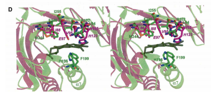
4. Vorobiev SM; Huang YJ; Seetharaman J; Xiao R; Acton TB; Montelione GT; Tong L. Protein Peptide Letts. 2011, 19: 194 – 197. Human retinoblastoma binding protein 9, a serine hydrolase implicated in pancreatic cancers. PMC3677193. Pubmed.
5. Acton TB; Xiao R; Anderson S; Aramini JM; Buchwald W; Ciccosanti C; Conover K; Everett JK; Hamilton K; Huang YJ; Janjua H; Kornhaber GJ; Lau J; Lee DY; Liu G; Maglaqui M; Ma LC; Mao L; Patel D; Rossi P; Sahdev S; Sharma S; Shastry R; Swapna GVT; Tang Y; Tong SN; Wang D; Wang H; Zhao L; Montelione GT. Methods in Enzymology. 2011, 493: 21 – 60. Preparation of protein samples for NMR structure, function, and small molecule screening studies. PMC4110644. Pubmed.
6. Mao L; Inoue K; Tao Y; Montelione GT; McDermott AE; Inouye M. J Biomol NMR. 2011, 49: 131 – 137. Suppression of phospholipid biosynthesis by cerulenin in the condensed Single-Protein- Production (cSPP) system. PMC3164850.
7. Ramelot TA; Smola MJ; Lee HW; Ciccosanti C; Hamilton K; Acton TB; Xiao R; Everett JK; Prestegard JH; Montelione GT; Kennedy MA. Biochemistry. 2011, 50: 1442 – 1453. Solution NMR structure of 4’-phosphopantetheine – GmACP3 from Geobacter metallireducens : a specialized acyl carrier protein with atypical structural features and a putative role in lipopolysaccharide biosynthesis. suppl. material. PMC3063093. Pubmed.
8. Yin C; Aramini JM; Ma LC; Cort JR; Swapna GVT; Krug RM; Montelione GT. J Biomol NMR Assignments. 2011 5: 215 – 219. Backbone and Ile-δ1, Leu, Val methy 1H, 13C and 15N NMR chemical shift assignments for human interferon-stimulated gene 15 protein. PMC3167004. Pubmed.
9. Karanicolas J; Corn JE; Chen I; Joachimiak LA; Dym O; Peck SH; Albeck S; Unger T; Hu W; Liu G; Delbecq S; Montelione GT; Spiegel CP; Liu DR; Baker D. Molecular Cell. 2011, 42: 250 – 260. A de novo protein binding pair by computational design and directed evolution. suppl. material. PMC3102007. Pubmed.
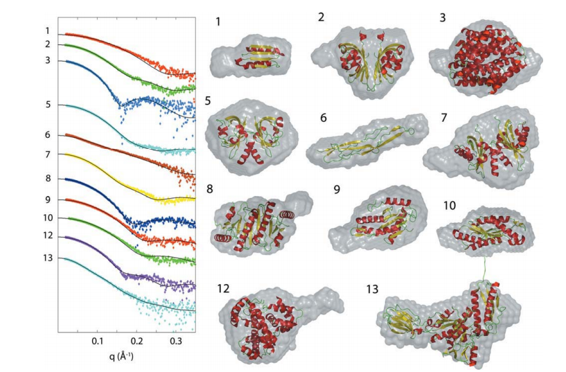
10. Grant T; Luft JR; Wolfley JR; Tsuruta H; Martel A; Montelione GT; Snell E. Biopolymers. 2011, 95: 517 – 530. Small angle x-ray scattering as a complementary tool for high-throughput structural studies. PMC3124082. Pubmed.
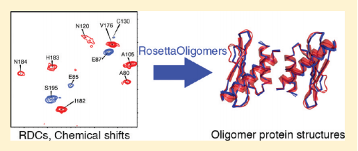
11. Sgourakis NG; Lange OF; DiMaio F; André I; Fitzkee NC; Rossi P; Montelione GT; Bax A; Baker D. J Amer Chem Soc. 2011, 133: 6288 – 6298. Determination of the structures of symmetric protein oligomers from NMR chemical shifts and residual dipolar couplings. suppl. material. PMC3080108. Pubmed.
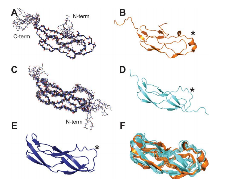
12. Barb AW; Cort JR; Seetharaman J; Lew S; Lee HW; Acton T; Xiao R; Kennedy MA; Tong L; Montelione GT; Prestegard JH. Protein Science. 2011, 20: 396 – 405. Structures of domains I and IV from YbbR are representative of a widely distributed protein family. PMC3048424. Pubmed.
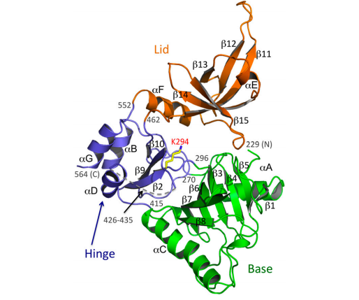
13. Chu C; Das K; Tyminski JR; Bauman JD; Guan R; Qiu W; Montelione GT; Arnold E; Shatkin AJ. Proc Natl Acad Sci USA. 2011, 108: 10104 – 10108. Structure of the guanylyltransferase domain of human mRNA capping enzyme. suppl. material. PMC3121809. Pubmed.
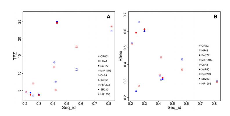
14. Mao B; Guan R; Montelione GT. Structure (Cell Press). 2011, 19: 757 – 766. Improved technologies now routinely provide protein NMR structures useful for molecular replacement. suppl. material. PMC3612016.
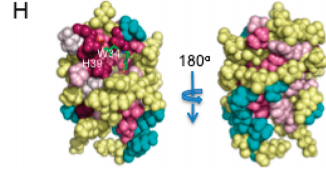
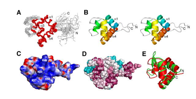
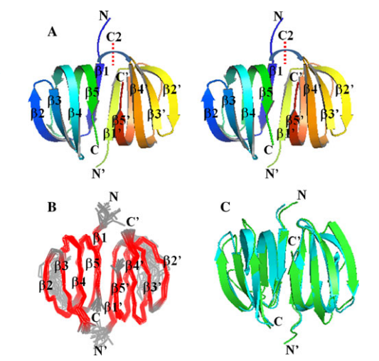
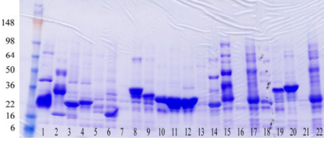
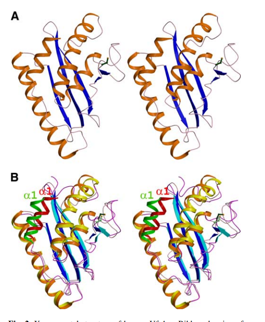
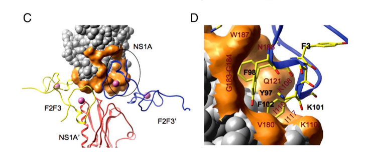
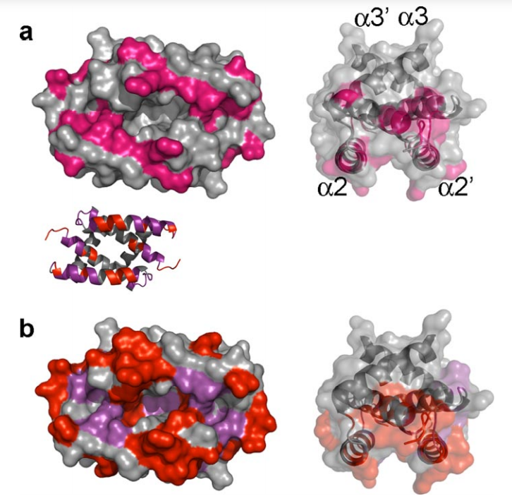
15. Aramini JM; Ma LC; Zhou L; Schauder CM; Hamilton K; Amer BR; Mack TR; Lee HW; Ciccosanti CT; Zhao L; Xiao R; Krug RM; Montelione GT. J Biol Chem. 2011, 286: 26050 – 26060. The dimer interface of the effector domain of non-structural protein 1 from influenza A virus: an interface with multiple functions. suppl. material. PMC3138300. Pubmed.
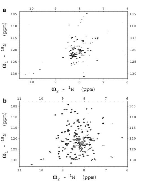
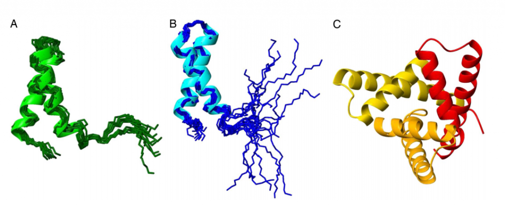
![Solution NMR studies of [W187R]Ud and wild type Ud NS1A(85–215)](https://montelionelab.chem.rpi.edu/wp-content/uploads/2025/07/image-56.png)
16. Aramini JM; Rossi P; Fischer M; Xiao R; Acton TB; Montelione GT. PROTEINS: Struct Funct Bioinformatics. 2011, 79: 2988 – 2991. Solution NMR structure of VF0530 from Vibrio fischeri reveals a nucleic-acid binding function. suppl. material. PMC3172673. Pubmed.
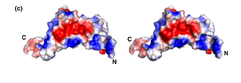
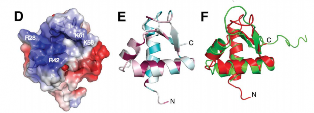
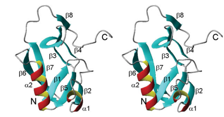
17. Guan R; Ma LC; Leonard PG; Amer BR; Sridharan H; Zhao C; Krug RM; Montelione GT. Proc Natl Acad Sci USA. 2011, 108: 13468 – 13473. Structural basis for the sequence-specific recognition of human ISG15 by the NS1 protein of influenza B virus. suppl. material. PMC3158222. Pubmed.

18. Eletsky A; Ruyechan WT; Xiao R; Acton TB; Montelione GT; Szyperski T. J Struct Funct Genomics. 2011, 12: 159 – 166. Solution NMR structure of MED25(391-543) comprising the activator-interacting domain (ACID) of human mediator subunit 25. suppl. material. PMC3609412. Pubmed.
19. Forouhar F; Saadat N; Hussain M; Seetharaman J; Lee I; Janjua H; Xiao R; Shastry R; Acton T; Montelione GT; Tong L. Acta Crystallographica Section F. 2011, 67: 1323 – 1327. A large conformational change in the putative ATP pyrophosphatase PF0828 induced by ATP binding. PMC3212444. Pubmed.
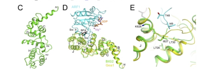
20. Lowery J; Szul T; Seetharaman J; Xiaoying J; Su M; Forouhar F; Xiao R; Acton TB; Montelione GT; Lin H; Wright JW; Lee E; Holloway ZG; Randazzo PA; Tong L; Sztul E. J Biol Chem. 2011, 286: 36898 – 36906. A novel C-terminal motif within the Sec7 domain of guanine nucleotide exchange factors regulates ARF binding and activation. suppl. material. PMC3196086. Pubmed.
21. Yang Y; Ramelot TA; Cort JR; Wang D; Ciccosanti C; Jiang M; Acton TB; Xiao R; Everett JK; Montelione GT; Kennedy M. J Struct Funct Genomics. 2011, 12: 175 – 179. Solution NMR structure of Dsy0195 homodimer from Desulfitobacterium hafniense: First structure representative of the YapB domain family of proteins involved in spore coat assembly. suppl. material. PMC3697068.
22. Kryshtafovych, A.; Bartual, S.G.; Bazan, J.F.; Berman, H.; Casteel, D.E.; Christodoulou, E.; Everett, J.K.; Hausmann, J.; Heidebrecht, T.; Hills, T.; Hui, R.; Hunt, J.F.; Tong, L.; Seetharaman, J.; Joachimiak, A.; Kennedy, M.; Kim, C.; Lingel, A.; Michalska,K.; Montelione, G.T.; Otero, J.M.; Perrakis, A.; Pizarro, M.J.; van Raaij, M.J.; Ramelot, T.A.; Rousseau, F.; Weraimont, A.K.; Young, J.; Schwede, T. PROTEINS: Struct Funct Bioinformatics 2011, 79: S10: 6 – 20. Target highlights in CASP9: experimental target structures for the critical assessment of techniques from protein structure prediction. PMC369200. Pubmed.
23. Feldmann EA; Ramelot TA; Yang Y; Xiao R; Acton TB; Everett JK; Montelione GT; Kennedy MA. PROTEINS: Struct Funct Bioinformatics. 2011, 80: 671 – 675. Solution NMR structure of Asl3597 from Nostoc sp. PCC7120, the first structure from protein domain family PF12095, adopts a novel fold. suppl. material. PMC3315617. Pubmed.
2010
1. Singarapu KK; Mills J; Xiao R; Acton T; Punta M; Fischer M; Honig B; Rost B; Montelione GT; Szyperski T. PROTEINS: Struct Funct Bioinformatics. 2010, 78: 779 – 784. Solution NMR structures of proteins VPA0419 from Vibrio parahaemolyticus and yiiS from Shigella flexneri provide structural coverage from protein domain family PFAM 04175. suppl. material. PMC2860719. Pubmed.
2. Mao L; Vaiphei ST; Shimazu T; Schneider WM; Tang Y; Mani R; Roth MJ; Montelione GT; Inouye M. J Struct Funct Genomics. 2010, 11: 81-84. The E. coli single protein production (cSPP) system for production and structural analysis of membrane proteins. PMC4190415.
3. Rossi, P.; Swapna, G.V.T.; Huang, Y.J.; Aramini, J.M.; Anklin, C.; Conover, K.; Hamilton, K.; Xiao, R.; Acton, T.B.; Ertekin, A.; Everett, J.K.; Montelione, G.T. J Biomol NMR 2010, 46: 11 – 22. A microscale protein NMR sample screening pipeline. suppl. material PMC2797623. Pubmed.
4. Raman, S.; Huang, Y.J.; Mao, B.; Rossi, P.; Aramini, J.M.; Liu, G.; Montelione, G.T.; Baker, D. J Amer Chem Soc 2010, 132: 202 – 207. Accurate automated protein NMR structure determination using unassigned NOESY data. suppl. material PMC2841443. Pubmed.
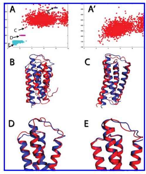
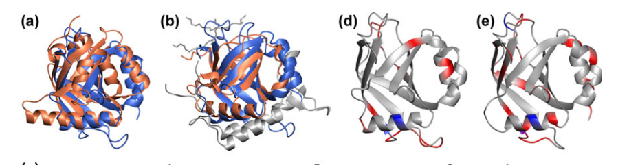
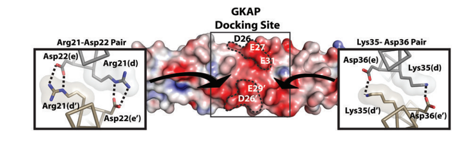
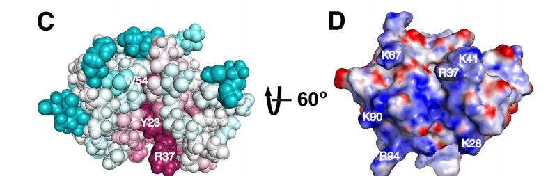
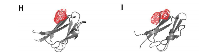
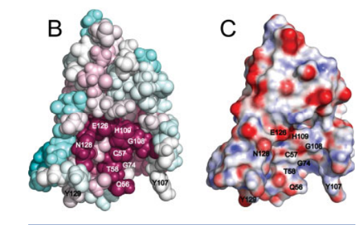
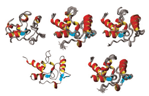
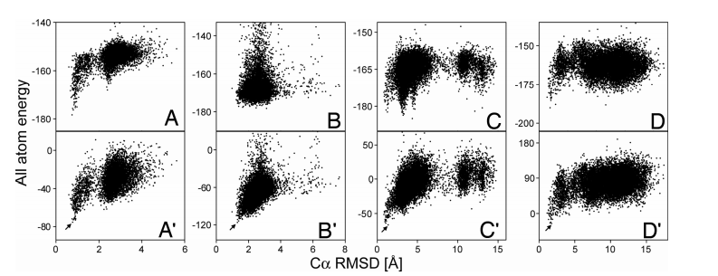
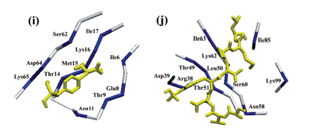
5. Raman, S.; Lange, O.F.; Rossi, P.; Tyka, M.; Wang, X.; Aramini, J.; Liu, G.; Ramelot, T.; Eletsky, A.; Szyperski, T.; Kennedy, M.; Prestegard, J.; Montelione, G.T.; Baker, D. Science 2010, 327: 1014 – 1018. NMR structure determination for larger proteins using backbone-only data. suppl. material PMC2909653. Pubmed.
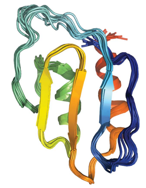
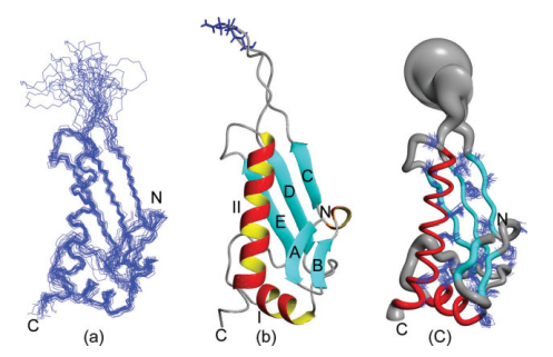
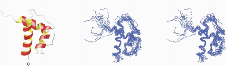
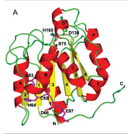
![Ensemble of 10 lowest-energy Rosetta structures [below line in (A)].
Regions with more than 3 Å RMSF are depicted in gray.](https://montelionelab.chem.rpi.edu/wp-content/uploads/2021/01/image-4.png)
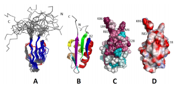
6. Liu, G.; Huang, Y.J.; Xiao, R.; Wang, D.; Acton, T.B.; Montelione, G.T. PROTEINS: Struct Funct Bioinformatics 2010, 78: 1326 – 1330. NMR structure F-actin binding domain of Arg/Ab12 from Homo sapiens. suppl. material PMC2821974. Pubmed.
7. Montelione, G.T.; Szyperski, T.; Curr Opin Drug Discovery 2010, 13: 335 – 349. Advances in protein NMR impacting drug discovery provided by the NIGMS Protein Structure Initiative. PMC4002360. Pubmed.

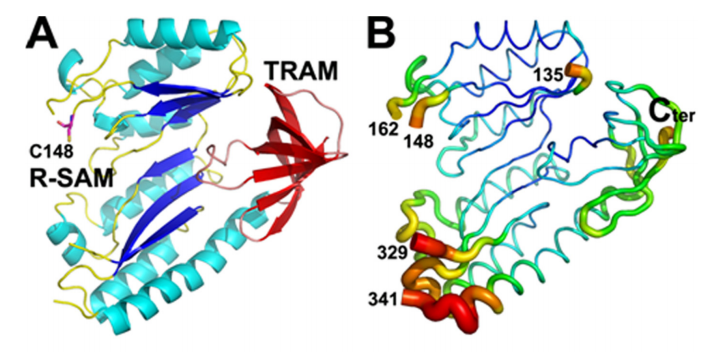
8. Arragain, S.; Garcia-Serres, R.; Blondin, G.; Douki, T.; Clemancey, M.; Latour, J-M.; Forouhar, F.; Neely, H.; Montelione, G.T.; Hunt, J.F.; Mulliez, E.; Fontecave, M.; Atta, M. J Biol Chem 2010, 285: 5792 – 5801. Post- translational modification of ribosomal proteins: Structural and functional characterization of RimO from Thermotoga maritima, a radical S- adenosylmethionine methylthiotransferase. suppl. material PMC282080. Pubmed.
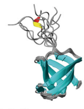
9. Aramini, J.M.; Tubbs, J.L.; Kanugula, S.; Rossi, P.; Ertekin, A.; Maglaqui, M.; Hamilton, K.; Ciccosanti, C.T.; Jiang, M.; Xiao, R.; Soong, T.- T.; Rost, B.; Acton, T.B.; Everett, J.K.; Pegg, A.E.; Tainer, J.A.; Montelione, G.T. J Biol Chem 2010, 285: 13736 – 13741. Structural basis of O6-alkylguanine recognition by a bacterial alkyltransferase-like DNA repair protein. suppl. material PMC2859536. Pubmed.
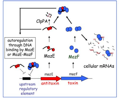
10. Arbing, M.A.; Handelman, S.K.; Kuzin, A.P.; Verdon, G.; Wang, C.; Su, M.; Rothenbacher, F.P.; Abashidze, M.; Liu, M.; Hurley, J.M.; Xiao, R.; Acton, T.; Inouye, M.; Montelione, G.T.; Woychik, N.A.; Hunt, J.F. Structure (Cell Press) 2010, 18: 996 – 1010. Crystal structures of Phd-Doc, HigA, and YeeU establish multiple evolutionary links between microbial growth-regulating toxin-antitoxin systems. suppl. material Open access journal. Pubmed.
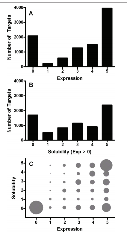
11. Price, W.N. 2nd; Handelman, S.K.; Everett, J.; Tong, S.N.; Bracic, A.; Luff, J.D.; Naumov, V.; Acton, T.; Manor, P.; Xiao, R.; Rost, B.; Montelione, G.T.; Hunt, J.F. Microbial Informatics and Experimentation 2011, 1: 6 – 26. Large-scale experimental studies show unexpected amino acid effects on protein expression and solubility in vivo in E. Coli. suppl. material PMC3372292. Pubmed.
12. Nie, Y.; Xiao, R.; Xu, Y.; Montelione, G.T. Org Biomol Chem 2011, 9: 4070 – 4078. Novel anti-Prelog stereospecific carbonyl reductases from Candida parapsilosis for asymmetric reduction of prochiral ketones. suppl. material PMC4104987. Pubmed.
13. Schneider, W.M.; Tang, Y.; Vaiphei, S.T.; Mao, L.; Maglaqui, M.; Inouye, M.; Roth, M.J.; Montelione, G.T. J Struct Funct Genomics 2010, 11: 143 – 154. Efficient condensed-phase production of perdeuterated soluble and membrane proteins. suppl. material PMC4119428. Pubmed.
14. Liu, G.; Huang, Y.J.; Xiao, R.; Wang, D.; Acton, T.B.; Montelione, G.T. PROTEINS: Struct Funct Bioinformatics 2010, 78: 2170 – 2175. Solution NMR structure of the ARID domain of human AT-rich interactive domain-containing protein 3A: A human cancer protein interaction network target. suppl. material PMC2869213. Pubmed.

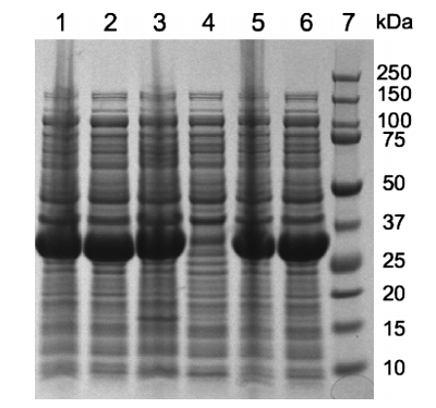
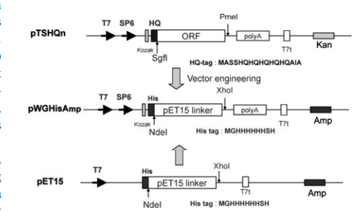
15. Zhao, L.; Zhao, K.; Hurst, R.; Slater, M.; Acton, T.B., Swapna, G.V.T.; Shastry, R.; Kornhaber, G.J.; Montelione, G.T. J Struct Funct Genomics 2010, 11: 210 – 209. Engineering of a wheat germ expression system to provide compatibility with a high throughput pET-based cloning platform. suppl. material PMC2921493. Pubmed.
16. Lee, H-W.; Wylie, G.; Bansal, S.; Wang, X.; Barb, A.W.; Macnaughtan, M.A.; Ertekin, A.; Montelione, G.T.; Prestegard, J.H. Protein Science 2010, 19: 1673 – 1685. Three-dimensional structures of the weakly associated protein homodimer SeR13 using RDCs and paramagnetic surface mapping. PMC2975131.
17. Stark, J.L.; Mercier, K.A.; Mueller, G.A.; Acton, T.B.; Xiao, R.; Montelione, G.T.; Powers, R. PROTEINS: Struct Funct Bioinformatics 2010, 78: 3328 – 3340. Solution structure and function of YndB, an AHSA1 protein from Bacillus subtilis. PMC2976784. Pubmed.
18. Tang, Y.; Xiao, R.; Ciccosanti, C.; Janjua, H.; Lee, Y.L.; Everett, J.; Swapna, G.V.T.; Acton, T.B.; Rost, B.; Montelione, G.T. PROTEINS: Struct Funct Bioinformatics 2010, 78: 2563 – 2568. Solution NMR structure of Lin0431 protein from Listeria innocua reveals high structural similarity with domain II of bacterial transcription antitermination protein NusG. suppl. material PMC2931792. Pubmed.
19. Xiao, R.; Anderson, S.; Aramini, J.M.; Belote, R.; Buchwald, W.; Ciccosanti, C.; Conover, K.; Everett, J.K.; Hamilton, K.; Huang, Y.J.; Janjua, H.; Jiang, M.; Kornhaber, G.J.; Lee, D.Y.; Locke, J.Y.; Ma, L.-C.; Maglaqui, M.; Mao, L.; Mitra, S.; Patel, D.; Rossi, P.; Sahdev, S.; Sharma, S.; Shastry, R.; Swapna, G.V.T.; Tong, S.N.; Wang, D.; Wang, H.; Zhao, L.; Montelione, G.T.; Acton, T.B. J Struct Biol 2010, 172: 21 – 33. The high- throughput protein sample production platform of the Northeast Structural Genomics Consortium. PMC4110633. Pubmed.
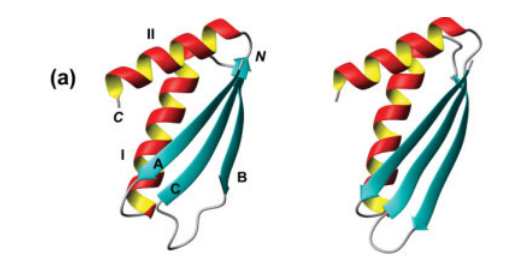
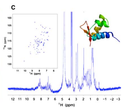
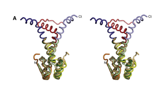
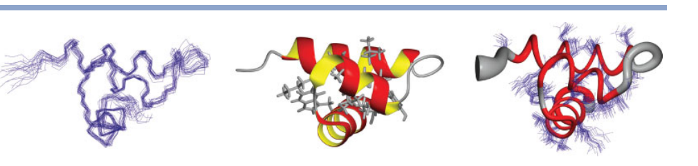
20. Tang, Y.; Schneider, W.M.; Shen, Y.; Raman, S.; Inouye, M.; Baker, D.; Roth, M.J.; Montelione, G.T. J Struct Funct Genomics 2010, 11: 223 – 232. Fully automated high quality NMR structure determination of small 2H- enriched proteins. PMC2970817.
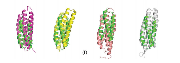
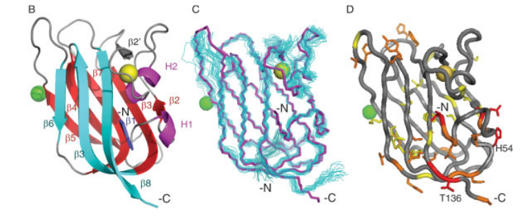
![Stereoview of the superimposition of AutoStructure-CNS structure for [1H-13C]-I(d1)LV, 13C, 15N, 2 H-enriched CspA determined by conventional automated analysis methods (blue) with 1mjc
(red).](https://montelionelab.chem.rpi.edu/wp-content/uploads/2021/01/image-18.png)
21. Yang, Y.; Ramelot, T.A.; Cort, J.R.; Wang, D.; Ciccosanti, C.; Hamilton, K.; Nair, R.; Rost, B.; Acton, T.B.; Xiao, R.; Everett, J.K.; Montelione, G.T.; Kennedy, M. PROTEINS: Struct Funct Bioinformatics 2011, 79: 340 – 344. Solution NMR structure of photosystem II reaction center protein Psb28 from Synechocystis sp. strain PCC 6803. suppl. material 1 suppl. material 2 suppl. material 3 PMC3248274. Pubmed.

22. Yang, Y.; Ramelot, T.A.; McCarrick, R.; Ni, S.; Feldmann, E.; Cort, J.R.; Wang, D.; Ciccosanti, C.; Jiang, M.; Janjua, H.; Acton, T.B.; Xiao, R.; Everett, J.K.; Montelione, G.T.; Kennedy, M. J Amer Chem Soc 2010, 132: 11910 – 11913. Combining NMR and EPR methods for homo-dimer protein structure determination. suppl. material. suppl. material. PMC3057626. Pubmed.
23. Love, J.; Mancia, F.; Shapiro, L.; Punta, M.; Rost, B.; Girvin, M.; Wang, D.-N.; Zhou, M.; Hunt, J.F.; Szyperski, T.; Gouaux, E.; MacKinnon, R.; McDermott, A.; Honig, B.; Inouye, M.; Montelione, G.T.; Hendrickson, W.A. J Struct Funct Genomics 2010, 11: 191 – 199. The New York Consortium on Membrane Structure (NYCOMPS): A high-throughput platform for structural genomics of integral membrane proteins. PMC3099345. Pubmed.
24. Mani, R.; Vorobiev, S.; Swapna, G.V.T.; Neely, H.; Janjua, H.; Ciccosanti, C.; Xiao, R.; Acton, T.B.; Everett, J.K.; Hunt, J.F.; Montelione, G.T. J Struct Funct Genomics 2011, 12: 27 – 32. Solution NMR and X-ray crystal structures of membrane-associated lipoprotein-17 domain reveal a novel fold. suppl. material PMC3636556
25. Zhang, H.; Constantine, R.; Vorobiev, S.; Chen, Y. ;Seetharaman, J.; Huang, Y.J.; Xiao, R.; Montelione, G.T.; Gerstner, C.D.; Davis, M.W.; Inana, G.; Whitby, F.G.; Jorgensen, E.M.; Hill, C.P.; Tong, L.; Baehr, W. Nature Neuroscience2011, 14: 874 – 880. UNC119 is required for G protein trafficking in sensory neurons. suppl. material PMC3178889. Pubmed.
26. Aramini, J.M.; Rossi, P.; Cort, J.; Ma, L.-C.; Xiao, R.; Acton, T.B.; Montelione, G.T. PROTEINS: Struct Funct Bioinformatics 2011, 79: 335 – 339. Solution NMR structure of the plasmid-encoded fimbriae regulatory protein PefI from Salmonella enterica serovar Typhimurium. suppl. material PMC2995844.
27. Forouhar, F.; Lew, S.; Seetharaman, J.; Xiao, R.; Acton, T.B.; Montelione, G.T.; Tong, L. Acta Cryst F 2010, F66: 1562 – 1566. Crystal structures of bacterial biosynthetic arginine decarboxylases. PMC2998355. Pubmed.
28. Montelione, G.T.; Szyperski, T. Advances in BioNMR Spectroscopy 2010, IOS Press, Editors A.J. Dingley and S.M. Pascal. Advances in NMR-based structural genomics spectroscopy.
2009
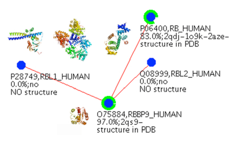
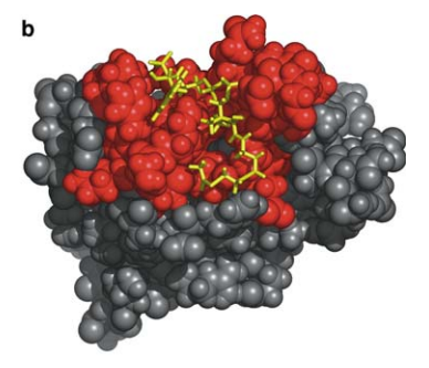
1. Vorobiev, S.M.; Su, M.; Seetharaman, J.; Huang, Y.J.; Chen, X.C.; Cunningham, K.; Maglaqui, M.; Owens, L.; Proudfoot, M.; Yakunin, A.; Xiao, R.; Acton, T.B.; Montelione, G.T.; Tong, L. PROTEINS: Struct Funct Bioinformatics 2009, 74: 526 – 529. Crystal structure of human retinoblastoma binding protein 9 (RBBP9). PMC2684859.
2. Cort, J.R.; Ramelot, T.A.; Murray, D.; Acton, T.B.; Ma, L-C.; Xiao, R.; Montelione, G.T.; Kennedy, M.A. PROTEINS: Struct Funct Bioinformatics 2008, 9: 7 – 20. Structure of an acetyl-CoA binding protein from Staphylococcus aureus representing a novel subfamily of GCN5- related N -acetyltransferase-like proteins.
3. Ramelot, T.A.; Raman, S.; Kuzin, A.P.; Xiao, R.; Ma, L.-C.; Acton, T.B.; Hunt, J.F.; Montelione, G.T.; Baker, D.; Kennedy, M.A. PROTEINS: Struct Funct Bioinformatics 2008, 75: 147 – 167. Improving NMR protein structure quality by Rosetta refinement: A molecular replacement study. suppl. material PMC2878636. Pubmed.

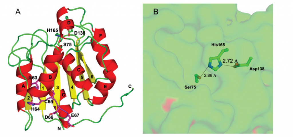
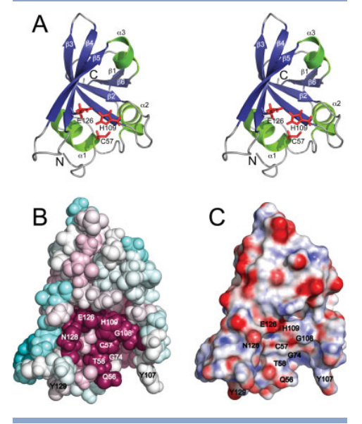
4. Rossi, P.; Aramini, J.M.; Xiao, R.; Chen, C.X.; Nwosu, C.; Owens, L.A.; Maglaqui, M.; Nair, R.; Fischer, M.; Acton, T.B.; Honig, B.; Rost, B.; Montelione, G.T. PROTEINS: Struct Funct Bioinformatics 2009, 74: 515 – 519. Structural elucidation of the Cys-His-Glu-Asn proteoytic relay in the secreted CHAP domain enzyme from the human pathogen Staphylococcus saprophyticus. suppl. material PMC2735724. Pubmed.
5. Parish D; Benach J; Liu G; Singarapu KK; Xiao R; Acton T; Su M; Bansal S; Prestegard JH; Hunt J; Montelione GT; Szyperski T. J Struct Funct Genomics. 2008, 9: 41 – 49. Protein chaperones Q8ZP25_SALTY from Salmonella typhimurium and HYAE_ECOLI from Escherichia coli exhibit thioredoxin-like structures despite lack of canonical thioredoxin active site sequence motif. PMC2850599.
6. Price WN. 2nd; Chen Y; Handelman SK; Neely H; Manor P; Karlin R; Nair R; Liu R; Baran M; Everett J; Tong SN; Forouhar F; Swaminathan SS; Acton T; Xiao R; Luft JR; Lauricella A; DeTitta GT; Rost B; Montelione GT; Hunt JF. Nature Biotechnology. 2009, 27: 51 – 57. Understanding the physical properties that control protein crystallization by analysis of large-scale experimental data. suppl. material. PMC2746436. Pubmed.
7. Liu G; Forouhar F; Eletsky A; Atreya HS; Aramini JM; Xiao R; Huang YJ; Abashidze M; Seetharaman J; Liu J; Rost B; Acton T.; Montelione GT; Hunt JF; SzyperskiT. J Struct Funct Genomics. 2009, 10: 127 – 136. NMR and X-ray structures of human E2-like ubiquitin-fold modifier conjugating enzyme 1 (UFC1) reveal structural and functional conservation in the metazoan UFM1-UBA5-UFC1 ubiquitination pathway. PMC2850604. Pubmed.
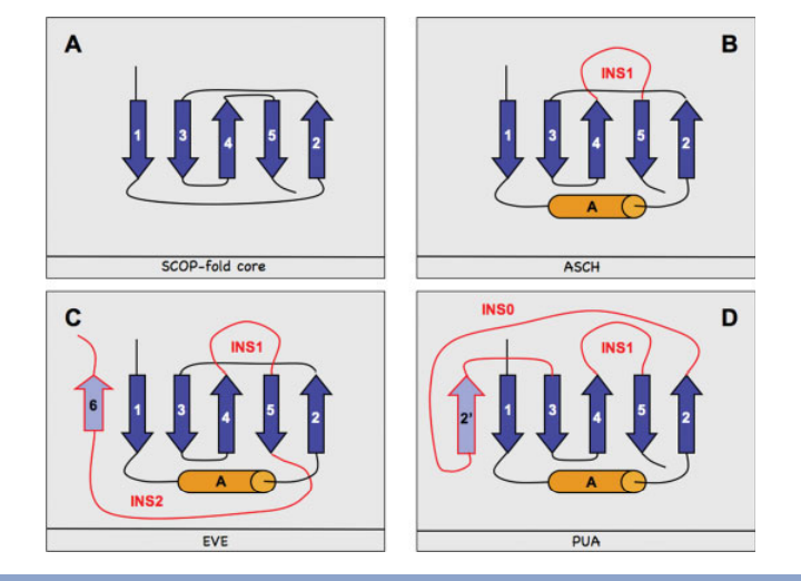
8. Bertonati C; Punta M; Fischer M; Yachdav G; Forouhar F; Zhou W; Kuzin AP; Seetharaman J; Abashidze M; Ramelot TA; Kennedy MA. PROTEINS: Struct Funct Bioinformatics. 2009, 75: 760 – 773. Structural genomics reveals EVE as a new ASCH/PUA-related domain. suppl. material. PMC4080787.
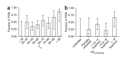
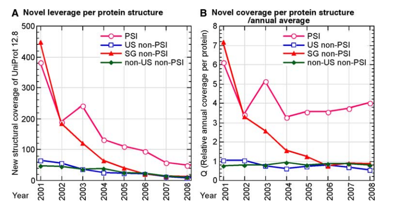
9. Nair R; Liu J; Soong TT; Acton TB; Everett J; Kouranov A; Fiser A; Godzik A; Jaroszewski L; Orengo C; Montelione GT; Rost B. J Struct Funct Genomics. 2009, 10: 181 – 191. Structural genomics is the largest contributor of novel structural leverage. PMC2705706. Pubmed.
10. Schwede T; Sali A; Honig B; Levitt M; Berman HM; Jones D; Brenner SE; Burley SK; Das R; Dokholyan NV; Dunbrack RL; Fidelis K; Fiser A; Godzik A; Huang YJ; Humblet C; Jacobson MP; Joachimiak A; Krystek SR Jr; Kortemme T; Kryshtafovych A; Montelione GT; Moult J; Murray D; Sanchez R; Sosnick TR; Standley DM; Stouch T; Vajda S; Vasquez M; Westbrook JD; Wilson IA. Structure (Cell Press). 2009, 17: 151 – 159. Outcome of workshop on applications of protein models in biomedical research. PMC2739730. Pubmed.
11. Montelione GT; Arrowsmith CA; Girvin M; Kennedy MA; Markley JL; Powers R; Prestegard JH; Szyperski T. J Struct Funct Genomics. 2009, 10: 101-106. Unique opportunities for NMR methods in structural genomics. PMC2705713. Pubmed.
12. Sharma S; Zheng H; Huang YJ; Ertekin A; Hamuro Y; Rossi P; Tejero R; Acton T; Xiao R; Jiang M; Zhao L; Ma LC; Swapna GVT; Aramini JM; Montelione GT. PROTEINS: Struct Funct Bioinformatics. 2009, 76: 882 – 894. Construct optimization for protein NMR structure analysis using amide hydrogen/deuterium exchange mass spectrometry. suppl. material. PMC2739808.
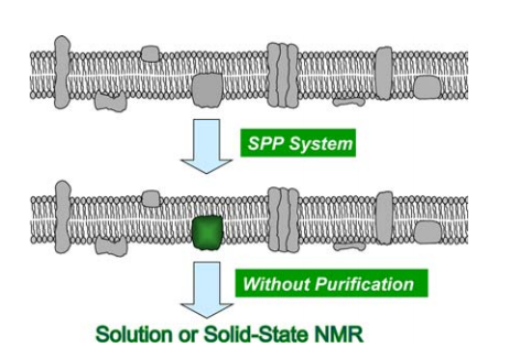
13. Mao L; Tang Y; Vaiphei T; Shimazu T; Kim SG; Mani R; Fakhoury E; White E; Montelione GT; Inouye M. J Struct Funct Genomics. 2009, 10: 281 – 289. Production of membrane proteins for NMR studies using the condensed single protein (cSPP) production system. suppl. material 1. suppl. material 2. PMC2923930. Pubmed.
14. Eletsky A; Sukumaran DK; Xiao R; Acton T; Rost B; Montelione GT; Szyperski T. PROTEINS: Struct Funct Bioinformatics. 2009, 76: 1037 – 1041. NMR structure of protein YvyC from Bacillus subtilis reveals unexpected structural similarity between two PFAM families. suppl. material. PMC2735722. Pubmed.
15. Schneider WM; Inouye M; Montelione GT; Roth MJ. J Struct Funct Genomics. 2009, 10: 219 – 225. Independently inducible system of gene expression for condensed single protein production (cSPP) suitable for high efficiency isotope enrichment. PMC4898478.
16. Rosato A; Bagaria A; Baker D; Bardiaux B; Cavalli A; Doreleijers JF; Giachetti A; Guerry P; Güntert P; Herrmann T; Huang YJ; Jonker HRA; Mao B; Malliavin TE; Montelione GT; Nilges M; Raman S; van der Schot G; Vranken WF; Vuister GW; Bonvin AMJJ. Nature Methods. 2009, 6: 625 – 626. CASD-NMR: Critical assessment of automated structure determination by NMR. PMC2841015. Pubmed.
17. Mercier KA; Mueller GA; Acton TB; Xiao R; Montelione GT; Powers R. J Biomol NMR Assignments. 2009, 3: 191 – 194. 1H, 13C, and 15N NMR assignments for the Bacillus subtilis yndB START domain. PMC4991356.
2008
1. Burley SK; Joachimiak A; Montelione GT; Wilson IA. Structure (Cell Press). 2008, 16: 5 – 11. Contributions to the NIH-NIGMS Protein Structure Initiative from the PSI production centers. PMC2678832. Pubmed.
2. Singarapu KK; Xiao R; Sukumaran DK; Acton T; Montelione GT; Szyperski T. PROTEINS: Struct Funct Bioinformatics. 2008, 70: 1650 – 1654. NMR structure of protein Cgl2762 from Corynebacterium glutamicum implicated in DNA transposition reveals a helix-turn-helix motif attached to a flexibly disordered leucine zipper. suppl. material. Pubmed.
3. Gräslund S; Nordlund P; Weigelt J; Bray J; Gileadi O; Knapp S; Oppermann U; Arrowsmith C; Hui R; Ming J; Park HW; Savchenko A; Yee A; Edwards A; Vincentelli R; Cambillau C; Kim R; Kim SH; Rao Z; Shi Y; Terwilliger TC; Kim CY; Hung LW; Waldo GS; Peleg Y; Albeck S; Unger T; Dym O; Prilusky J; Sussman JL; Stevens RC; Lesley SA; Wilson IA; Joachimiak A; Collart F; Dementieva I; Donnelly MI; Eschenfeldt WH; Kim Y; Stols L; Wu R; Zhou M; Burley SK; Emtage JS; Sauder JM; Thompson D; Bain K; Luz J; Gheyi T; Zhang F; Atwell S; Almo SC; Bonanno JB; Fiser A.; Swaminathan S; Studier FW; Chance MR; Sali A; Acton TB; Xiao R; Zhao L; Ma LC; Hunt JF; Tong L; Cunningham K; Inouye M; Anderson S; Janjua H; Shastry R; Ho C.K.; Wang, H; Jiang M; Montelione GT; Stuart DI; Owens RJ; Daenke S; Schütz A; Heinemann U; Yokoyama S; Büssow K; Gunsalus KC. Nature Methods. 2008, 5: 135 – 146. Protein production and purification. PMC3178102. Pubmed.
4. Singarapu KK; Xiao R; Acton T; Rost B; Montelione GT; Szyperski T. PROTEINS: Struct Funct Bioinformatics. 2008, 71: 1027 – 1031. NMR structure of the peptidyl-tRNA hydrolase domain from Pseudomonas syringae expands the structural coverage of the hydrolysis domains of class 1 peptide chain release factors. Pubmed.
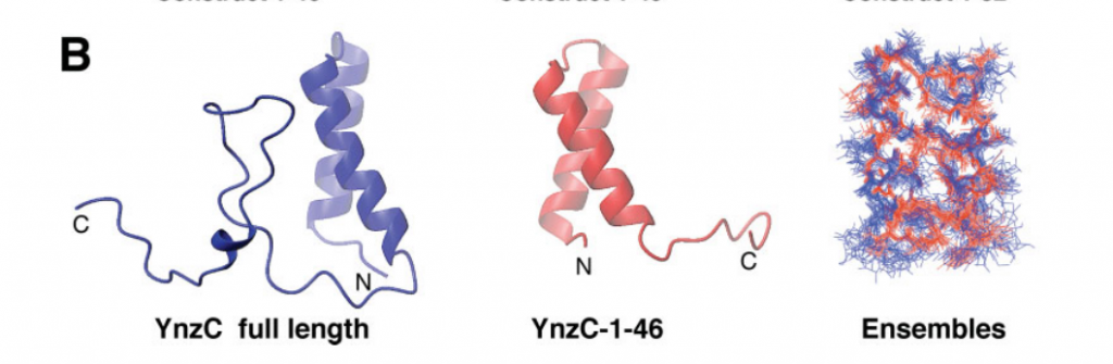
5. Aramini JM; Sharma S; Huang YJ; Swapna GVT; Ho CK; Shetty K; Cunningham K; Ma LC; Zhao L; Owens LA; Jiang M; Xiao R; Liu J; Baran MC; Acton TB; Rost B; Montelione GT. PROTEINS: Struct Funct Bioinformatics. 2008, 72: 526 – 530. Solution NMR structure of the SOS response protein YnzC from Bacillus subtilis. suppl. material. Pubmed.
6. Forouhar F; Abashidze M; Xu H; Grochowski LL; Seetharaman J; Hussain M; Kuzin A; Chen Y; Zhou W; Xiao R; Acton TB; Montelione GT; Galinier A; White RH; Tong L. J Biol Chem. 2008, 17: 11832 – 11840. Molecular insights into the biosythesis of the F420 coenzyme. PMC2431047. Pubmed.
7. ShenY; Lange O; Delaglio F; Rossi P; Aramini JM; Liu G; Eletsky A; Wu Y; Singarapu KK; Lemak A; Ignatchenko A; Arrowsmith CH; Szyperski T; Montelione GT; Baker D; Bax A. Proc Natl Acad Sci USA. 2008, 105: 4685 – 4690. Consistent blind protein structure generation from NMR chemical shift data. suppl. material. PMC2290745. Pubmed.
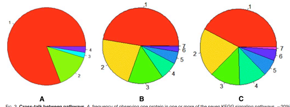
8. Huang YJ; Hang D; Lu LJ; Tong L; Gerstein MB; Montelione GT. Mol. Cell. Proteomics. 2008, 7: 2048 – 2060. Targeting the human cancer pathway protein interaction network by structural genomics. PMC2559933. Pubmed.
9. Wu B; Yee A; Huang YJ; Ramelot TA; Cort JR; Semesi A; Jung JW; Lee W; Montelione GT; Kennedy MA; Arrowsmith CH. Protein Science. 2008, 17: 583 – 588. The solution structure of ribosomal protein S17E from Methanobacterium thermoautotrophicum: A structural homolog of the FF domain. PMC2248302.
10. Das K; Ma LC; Xiao R; Radvansky B; Aramini J; Zhao L; Marklund J; Kuo RL; Twu KY; Arnold E; Krug RM; Montelione GT. Proc Natl Acad Sci USA. 2008, 105: 13092 – 13097. Structural basis for suppression by influenza A virus of a host antiviral response. suppl. material. PMC2522260. Pubmed.
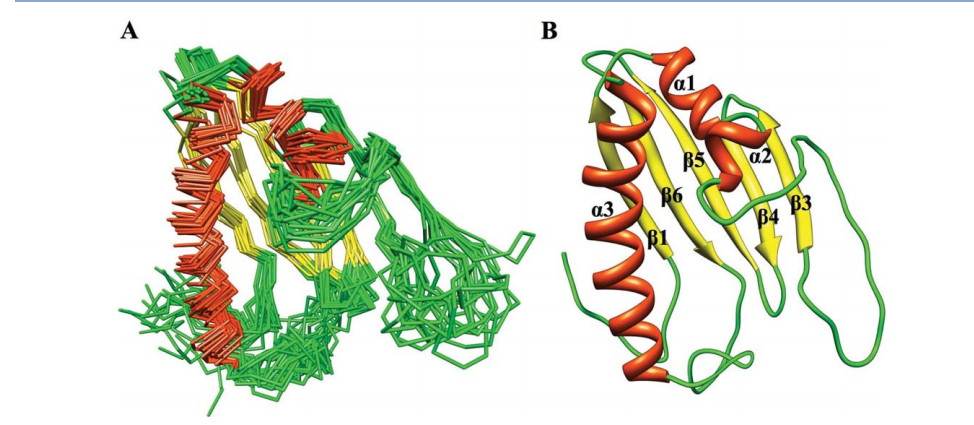
11. Aramini JM; Rossi P; Huang YJ; Zhao L; Jiang M; Maglaqui M; Xiao R; Locke J; Nair R; Rost B; Acton TB; Inouye M; Montelione GT. Biochemistry (Rapid Report). 2008, 47: 9715 – 9717. Solution NMR structure of the NIpC/P60 domain of lipoprotein Spr from Escherichia coli: Structural evidence for a novel cysteine peptidase catalytic triad. suppl. material. Pubmed.
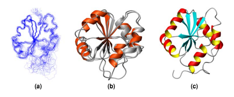
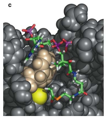
![View into the active site of E. coli Spr[37-162] showing the conserved Cys-His-His catalytic triad and flanking tyrosine. Juxtaposed heavy atoms in the triad and secondary structural elements are labeled.](https://montelionelab.chem.rpi.edu/wp-content/uploads/2021/01/image-48.png)
12. Vila J; Aramini J; Rossi P; Kuzin A; Su M; Seetharaman J; Xiao R; Tong L; Montelione GT; Scheraga H. Proc Natl Acad Sci USA. 2008, 105: 14389 – 14394. Quantum chemical 13Cα chemical shift calculations for protein NMR structure determination, refinement, and validation. suppl. material. PMC2567219.
13. Vorobiev SM; Su M; Seetharaman J; Huang YJ; Chen XC; Cunningham K; Maglaqui M; Owens L; Proudfoot M; Yakunin A; Xiao R; Acton TB; Montelione GT; Tong L. PROTEINS: Struct Funct Bioinformatics. 2009, 74: 526 – 529. Crystal structure of human retinoblastoma binding protein 9 (RBBP9). PMC2684859.
14. Cort JR; Ramelot TA; Murray D; Acton TB; Ma LC; Xiao R; Montelione GT; Kennedy MA. PROTEINS: Struct Funct Bioinformatics. 2008, 9: 7 – 20. Structure of an acetyl-CoA binding protein from Staphylococcus aureus representing a novel subfamily of GCN5- related N -acetyltransferase-like proteins.
15. Ramelot TA; Raman S; Kuzin AP; Xiao R; Ma LC; Acton TB; Hunt JF; Montelione GT; Baker D; Kennedy MA. PROTEINS: Struct Funct Bioinformatics. 2008, 75: 147 – 167. Improving NMR protein structure quality by Rosetta refinement: A molecular replacement study. suppl. material. PMC2878636. Pubmed.
16. Rossi P; Aramini JM; Xiao R; Chen CX; Nwosu C; Owens LA; Maglaqui M; Nair R; Fischer M; Acton TB; Honig B; Rost B; Montelione GT. PROTEINS: Struct Funct Bioinformatics. 2009, 74: 515 – 519. Structural elucidation of the Cys-His-Glu-Asn proteoytic relay in the secreted CHAP domain enzyme from the human pathogen Staphylococcus saprophyticus. suppl. material. PMC2735724. Pubmed.
17. Parish D; Benach J; Liu G; Singarapu KK; Xiao R; Acton T; Su M; Bansal S; Prestegard JH; Hunt J; Montelione GT; Szyperski T. J Struct Funct.Genomics. 2008, 9: 41 – 49. Protein chaperones Q8ZP25_SALTY from Salmonella typhimurium and HYAE_ECOLI from Escherichia coli exhibit thioredoxin-like structures despite lack of canonical thioredoxin active site sequence motif. PMC2850599.

2007
1. Mercier KA; Baran M; Ramanathan V; Revesz P; Xiao R; Montelione GT; Powers R. J Amer Chem Soc. 2006, 128: 15292 – 15299. FAST-NMR – Functional Annotation Screening Technology using NMR spectroscopy. PMC2529462. Pubmed.
2. Bhattacharya A; Tejero R; Montelione GT. PROTEINS: Struct Funct Bioinformatics. 2007, 66: 778 – 795. Evaluating protein structures determined by structural genomics consortia. Pubmed.
3. Forouhar F; Anderson JL; Mowat CG; Vorobiev SM; Hussain A; Abashidze M; Bruckmann C; Thackray SJ; Seetharaman J; Tucker T; Xiao R; Ma L; Zhao L; Acton TB; Montelione GT; Chapman SK; Tong L. Proc Natl Acad Sci USA. 2007, 104: 473 – 478. Molecular insights into substrate recognition and catalysis by tryptophan 2,3- dioxygenase. suppl. material. Pubmed.
4. Vorobiev SM; Neely H; Seetharaman J; Ma L; Xiao R; Acton TB; Montelione GT; Tong L. Protein Science. 2007, 16: 535 – 538. Crystal structure of AGR_C_4470p from Agrobacterium tumefaciens. PMC2203313. Pubmed.
5. Singarapu KK; Liu G; Xiao R; Bertonati C; Honig B; Montelione GT; Szyperski T. PROTEINS: Struct Funct Bioinformatics. 2007, 67: 501 – 504. NMR structure of protein yjbR from Escherichia coli reveals ‘double-wing’ DNA binding motif. Pubmed.
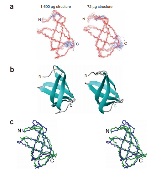
6. Aramini J; Rossi P; Anklin C; Xiao R; Montelione GT. Nature Methods. 2007, 4: 491 – 493. Microgram scale protein structure determination by NMR. suppl. material. Pubmed.
7. Yin C; Khan JA; Swapna GVT; Ertekin A; Krug RM; Tong L; Montelione GT. J Biol Chem. 2007, 282: 20584 – 20592. Conserved surface features form the double-stranded RNA-binding site of non-structural protein 1 (NS1) from Influenza A and B viruses. suppl. material. Pubmed.
8. Aramini J; Y. Huang YJ; Swapna GVT; Cort JR; Rajan PK; Xiao R; Shastry R; Acton TB; Liu J; Rost B; Kennedy MA; Montelione GT. PROTEINS: Struct Funct Bioinformatics. 2007, 68: 789 – 795. The solution NMR structure of Escherichia coli ytfP expands the structural coverage of the UPF0131 protein domain family. suppl. material. Pubmed.
9. Liu J; Montelione GT; Rost B. Nature Biotechology. 2007, 25: 849 – 851. Novel leverage of structural genomics. suppl. material. Pubmed.
10. Andrec M; Snyder D A; Zhou Z; Young J; Montelione GT; Levy R. PROTEINS: Struct Funct Bioinformatics. 2007, 69: 449 – 465. A large data set comparison of protein structures determined by crystallography and NMR: Statistical test for structural differences and the effect of crystal packing. suppl. material. Pubmed.
11. Lu LJ; Sboner A; Huang Y J; Lu H.X.; Gianoulis T.A.; Yip KY; Kim PM; Montelione GT; Gerstein MB. TIBS. 2007, 32: 320 – 331. Comparing classical pathways and modern networks: Towards the development of an edge ontology. suppl. material. Pubmed.
12. Bhattacharya A; Wunderlich Z; Monleon D; Tejero R; Montelione GT. PROTEINS: Struct Funct Bioinformatics. 2007, 70: 105 – 118. Assessing model accuracy using the homology modeling automatically (HOMA) software. suppl. material. Pubmed.
13. Forouhar F; Kuzin A; Seetharaman J; Lee I; Zhou W; Abashidze M; Chen Y; Yong W; Janjua H; Fang Y; Wang D; Cunningham K; Xiao R; Acton TB; Pichersky E; Klessig DF; Porter CW; Montelione GT; Tong L. J Struct Funct Genomics. 2007, 8: 37 – 44. Functional insights from structural genomics. Pubmed.
14. Benach J; Wang L; Chen Y; Ho CK; Lee S; Seetharaman J; Xiao R; Acton TB; Montelione GT; Deng H; Sun R; Tong L. J Biol Chem. 2007, 43: 31534 – 31541. Structural and functional studies of the abundant tegument protein ORF52 from murine gammaherpesvirus-68. Pubmed.

2006
1. Liu G; Shen Y; Xiao R; Acton T; Ma L; Joachimiak A; Montelione GT; Szyperski T. PROTEINS: Struct Funct Bioinformatics. 2006, 62: 288 – 291. NMR structure of protein yqbG from Bacillus subtilis reveals a novel α-helical protein fold. Pubmed.
2. Baran M; Moseley HNB; Aramini JM; Bayro MJ; Monleon D; Locke J; Montelione GT. PROTEINS: Struct Funct Bioinformatics. 2006, 62: 843 – 851. SPINS: A laboratory information management system for organizing and archiving intermediate and final results from NMR protein structure determinations. suppl. material. Pubmed.
3. Forouhar F; Hussain M; Farid R; Benach J; Abashidze M; Edstrom WC; Vorobiev SM; Xiao R; Acton TB; Fu Z; Kim J; Miziorko HM; Montelione GT; Hunt JF. J Biol Chem. 2006, 281: 7533 – 45. Crystal structures of two bacterial 3-hydroxy- 3methylglutaryl-CoA lyases suggest a common catalytic mechanism among a family of TIM-barrel metalloenzymes cleaving carbon-carbon bonds. Pubmed.
4. Kornhaber GJ; Snyder D; Moseley HNB; Montelione GT. J Biomol NMR. 2006, 34: 259 – 269. Identification of zinc-ligated cysteine residues based on 13Cα and 13Cβ chemical shift data. Pubmed.
5. Berman HM; Burley SK; Chiu W; Sali A; Adzhubei A; Bourne PE; Bryant SH; Dunbrack Jr, RL; Fidelis, K.; Frank, J.; Godzik, A.; Henrick, K.; Joachimiak, A.; Heymann, B.; Jones, D.; Markley, J.L.; Moult, J.; Montelione, G.T.; Orengo, C.; Rossman, M.T.; Rost, B.; Saibil, H.; Schwede, T.; Standley, D.M.; Westbrook, J.D. Structure (Cell Press) 2006, 14: 1211 – 1217. Outcome of a workshop on archiving structural models of biological macromolecules. Pubmed.
6. Greenfield NJ; Huang YJ; Swapna GVT; Bhattacharya A; Rapp B; Singh A; Montelione GT; Hitchcock-DeGregori SE. J Mol Biol. 2006, 364: 80 – 96. Solution NMR structure of the junction between tropomyosin molecules: Implications for actin binding and regulation. suppl. material. Pubmed.
7. Mercier KA; Baran M; Ramanathan V; Revesz P; Xiao R; Montelione GT; Powers R. J Amer Chem Soc. 2006, 128: 15292 – 15299. FAST-NMR – Functional Annotation Screening Technology using NMR spectroscopy. PMC2529462. Pubmed.

2005
1. Huang YJ; Moseley HNB; Baran MC; Arrowsmith C; Powers R; Tejero R; Szyperski T; Montelione GT. Methods in Enzymology. 2005, 394: 111 – 141. An integrated platform for automated analysis of protein NMR structures. Pubmed.
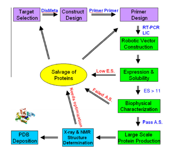
2. Acton TB; Gunsalus KC; Xiao R; Ma LC; Aramini JM; Baran MC; Chiang YW; Climent T; Cooper B; Denissova N; Douglas SM; Everett JK; Ho CK; Macapagal D; Rajan PK; Shastry R; Shih LY; Swapna GVT; Wilson M; Wu M; Gerstein M; Inouye M; Hunt JF; Montelione GT. Methods in Enzymology. 2005, 394: 210 – 243. Robotic cloning and protein production platform of the Northeast Structural Genomics Consortium. Pubmed.
3. Shen Y; Goldsmith-Fischman S; Atreya HS; Acton TB; Ma LC; Xiao R; Honig B; Montelione GT; Szyperski T. PROTEINS: Struct. Funct. Bioinformatics. 2005, 58: 747 – 750. NMR structure of the 18 kDa protein CC1736 from Caulobacter crescentus identifies a member of the “START” domain superfamily and suggests residues mediating substrate specificity.
4. Snyder DA; Montelione GT. PROTEINS: Struct Funct Bioinformatics. 2005, 59: 673 – 686. Clustering algorithms for identifying core atom sets and for assessing the precision of protein structure ensembles. Pubmed.
5. Huang YJ; Powers R; Montelione GT. J. Amer Chem Soc. 2005, 127: 1665 – 1674. Protein NMR Recall, Precision, and F-measure scores (RPF Scores): Structure quality assessment measures based on information retrieval statistics. suppl. material. Pubmed.
6. Liu G; Li Z; Chiang Y; Acton TB; Montelione GT; Murray D; Szyperski T. Protein Science. 2005, 14:1597 – 1608. High-quality homology derived from NMR and X-ray structures of E. coli. proteins YgdK and SufE suggest that all members of the YgdK/SufE protein family are enhancers of cysteine desulfurases. PMC2253389. Pubmed.
7. Forouhar F; Yang Y; Kumar D; Chen Y; Fridman E; Park SW; Chiang Y; Acton TB; Montelione GT; Pichersky E; Klessig DF; Tong L. Proc Natl Acad Sci USA. 2005, 102: 1773 – 1778. Structure and biochemical studies identify tobacco SABP2 as a methyl salicylate esterase and implicate it in plant innate immunity. suppl. material. PMC547883. Pubmed.
8. Liu G; Shen Y; Atreya HS; Parish D; Shao Y; Sukumaran DK; Xiao R; Yee A; Lemak A; Bhattacharya A; Acton TB; Arrowsmith CH; Montelione GT; Szyperski T. Proc Natl Acad Sci USA. 2005, 102: 10487 – 10492. NMR data collection and analysis protocol for high-throughput protein structure determination. suppl. material. PMC1180791. Pubmed.
9. Douglas SM; Montelione GT; Gerstein M. Genome Biology. 2005, 6:R80. PubNet: A flexible system for visualizing literature-derived networks. PMC1242215. Pubmed.
10. Huang YJ; Tejero R; Powers R; Montelione GT. PROTEINS: Struct Funct Bioinformatics. 2006, 62: 587 – 603. A topology- constrained distance network algorithm for protein structure determination from NOESY data. suppl. material. Pubmed.
11. Snyder DA; Bhattacharya A; Huang YJ.; Montelione GT. PROTEINS: Struct Funct Bioinformatics. 2005, 59: 655 – 661. Assessing precision and accuracy of protein structures derived from NMR data. suppl. material. Pubmed.
12. Snyder DA; Chen Y; Denissova NG; Acton T; Aramini JM; Ciano M; Karlin R; Liu J; Manor P; Rajan PA; Rossi P; Swapna GVT; Xiao R; Rost B; Hunt J; Montelione GT. J Amer Chem Soc. 2005, 127: 16505 – 16511. Comparisons of NMR spectral quality and success in crystallization demonstrate that NMR and x-ray crystallography are complementary methods for small protein structure determination. suppl. material. Pubmed.
13. Benach J; Edstrom WC; Lee I; Das K; Cooper B; Xiao R; Liu J; Rost B; Acton TB; Montelione GT; Hunt JF. Acta Cryst D: Biol Cryst. 2005, D61: 589 – 598. The 2.35 Å structure of the TenA homolog from Pyrococcus furiosus supports an enzymatic function in thiamine metabolism. Pubmed.
14. Rossi P; Ramelot T; Xiao R; Ho CK; Ma L; Acton TB; Kennedy MA; Montelione GT. J Biomol NMR. 2005, 33: 197. 1H, 13C, 15N resonance assignments for the protein coded by gene locus BB0938 of Bordetella bronchiseptica.
15. Aramini JM; Swapna GVT; Huang YJ; Rajan PK; Xiao R; Shastry R; Acton TB; Cort JR; Kennedy MA; Montelione GT. J Biomol NMR. 2005, 33: 197. 1H, 13C, 15N resonance assignments for Escherichia coli ytfP, a member of the broadly conserved UPF0131 protein domain family. Pubmed.
16. Liu G; Aramini J; Atreya HS; Eletsky A; Xiao R; Acton TB; Ma LC; Montelione GT; Szyperski T. J Biomol NMR. 2005, 32: 261. Letter to the Editor: GFT NMR based resonance assignment for the 21 kDa human protein UFC1. suppl. material. Pubmed.
17. Forouhar F; Lee IS; Vujcic J; Vujcic S; Shen J; Vorobiev SM; Xiao R; Acton TB; Montelione GT; Porter CW; Tong L. J Biol Chem. 2005, 280: 40328 – 40336. Structural and functional evidence for Bacillus subtilis PaiA as a novel N1-spermidine/spermine acetyltransferase (SSAT). Pubmed.
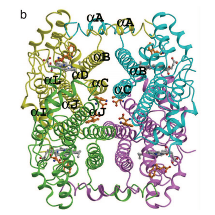
18. Bachhawat P; Swapna GVT; Montelione GT; Stock AM. Structure (Cell Press). 2005, 13: 1353 – 1363. Mechanism of activation for transcription factor PhoB suggested by different modes of dimerization in the inactive and active states. PMC3685586. Pubmed.
19. Powers R; Mirkovic N; Goldsmith-Fischman S; Acton TB; Chiang Y; Huang YJ; Ma L; Rajan RK; Cort JR; Kennedy MA; Liu J; Rost B; Honig B; Murray D; Montelione GT. Protein Science. 2005, 14: 2849 – 2861. Solution structure of Archaeglobus fulgidis peptidyl-tRNA hydrolase (Pth2) provides evidence for an extensive conserved family of Pth2 enzymes in archea, bacteria and eukaryotes. PMC2253226. Pubmed.
20. Huang YJ; Montelione GT. Nature. 2005, 438: 36 – 37. News and Views: Proteins flex to function. Pubmed.

2004
1. Monleon D; Chiang Y; Aramini JM; Swapna GVT; Macapagal D; Gunsalus KC; Kim S; Szyperski T; Montelione GT. J. Biomol. NMR. 2004, 28: 91 – 92. Backbone 1H, 15N and 13C assignments for the 21 kDa Caenorhabditis elegans homologue of ‘brain-specific’ protein. Pubmed.
2. Zheng D; Aramini J; Montelione GT. Protein Science. 2004, 13: 549 – 554. Validation of helical tilt angles in the solution NMR structure of the Z domain of Staphylococcal protein A by combined analysis of residual dipolar coupling and NOE data. PMC2286702.
3. Goh CS; Lan N; Douglas SM; Wu B; Echols N; Smith A; Milburn D; Montelione GT; Zhao H; Gerstein M. J. Mol. Biol. 2004, 336: 115 – 130. Mining the structural genomics pipeline: Identification and analysis of protein properties that affect high-throughput experimental analysis. Pubmed.
4. Chien CY; Xu Y; Xiao R; Aramini JM; Sahasrabudhe PV; Krug RM; Montelione GT. Biochemistry. 2004, 43: 1950 – 1962. Biophysical characterization of the complex between double-stranded RNA and the N-terminal domain of the NS1 protein from Influenza A virus: Evidence for a novel RNA-binding mode. Pubmed.
5. Moseley HNB; Sahota G; Montelione GT. J. Biomol. NMR. 2004, 28: 341 – 355. Assignment validation software suite for the evaluation and presentation of protein resonance assignment data. Pubmed.
6. Das K; Acton TB; Chiang Y; Shih L; Arnold E; Montelione GT. Proc. Natl. Acad. Sci. U.S.A. 2004, 101: 4041 – 4046. Crystal structure of E. coli RlmAI: Implications for understanding 23S rRNA G745/G748-methylation at the macrolide antibiotic-binding site. PMC384692. Pubmed.
7. Liu G; Sukumaran DK; Xu D; Chiang Y; Acton TB; Goldsmith- Fischman, S; Honig B; Montelione GT; Szyperski T. PROTEINS: Struct. Funct. Genetics. 2004, 55: 756 – 758. NMR structure of the hypothetical protein NMA1147 from Neisseria meningitidis reveals a distinct 5-helix bundle. Pubmed.
8. Herve Du Penhoat C; Atreya HS; Shen Y; Liu G; Acton TB; Xiao R; Li Z; Murray D; Montelione GT; Szyperski T. Protein Sci. 2004, 13: 1407 – 1416. The NMR solution structure of the 30S ribosomal protein S27e encoded in gene RS27_ARCFU of Archaeoglobus fulgidis reveals a novel protein fold. PMC2286747.
9. Xu D; Liu G; Xiao R; Acton T; Goldsmith-Fischman S; Honig B; Montelione GT; Szyperski T. PROTEINS: Struct. Funct. Genetics. 2004, 54: 794 – 796. NMR structure of the hypothetical protein AQ- 1857 encoded by the Y157 gene from Aquifex aeolicus reveals a novel protein fold. Pubmed.
10. Forouhar F; Lee IS; Benach J; Kulkarni K; Xiao R; Acton TB; Montelione GT; Tong L. J. Biol. Chem. 2004, 279: 13148 – 13155. A novel NAD binding protein revealed by the crystal structure of E. coli 2,3-diketogulonate reductase (YiaK). Pubmed.
11. Makokha M; Huang YJ; Montelione GT; Edison AS; Barbar E. Protein Science 2004 13: 727 – 734. The solution structure of the pH-induced monomer of dynein light-chain LC8 from Drosophila. PMC2286742. Pubmed.
12. Everett JK; Acton TB; Montelione G.T. J. Struct. Funct. Genomics. 2004, 5: 13 – 21. Primer Prim’ər: A web based server for automated primer design. Pubmed.
13. Adams M; Joachimiak A; Kim R; Montelione GT; Norvell J. J. Struct. Funct. Genomics. 2004, 5: 1 – 2. 2003 NIH protein structure initiative workshop in protein production and crystallization for structural and functional genomics (Meeting Review). Pubmed.
14. Wunderlich Z; Acton TB; Liu J; Kornhaber G; Everett J; Carter P; Lan N; Echols N; Gerstein M; Rost B; Montelione GT. PROTEINS: Struct. Funct. Bioinformatics. 2004, 56: 181 – 187. The protein target list of the Northeast Structural Genomics Consortium. Pubmed.
15. Shen Y; Atreya HS; Xiao R; Acton TB; Shastry R; Ma L; Montelione GT; Szyperski T. J. Biomol. NMR. 2004, 29: 549 – 550. Resonance assignments for the 18kDa protein CC1736 from Caulobacter crescentus.
16. Liu J; Hegyi H; Acton TB; Montelione GT; Rost B. PROTEINS: Struct. Funct. Bioinformatics. 2004, 56: 188 – 200. Automatic target selection for structural genomics on eukaryotes.
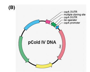
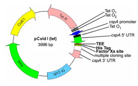
17. Qing G; Ma LC; Khorchid A; Swapna GVT; Mal TK; Takayama MM; Xia B; Phadtare S; Ke H; Acton T; Montelione GT.; Ikura M.; Inouye M. Nature Biotechnology. 2004, 22: 877 – 882. Cold-shock induced high-yield protein production in Escherichia coli. suppl. material. Pubmed.
18. Baran MC; Huang YJ; Moseley HN; Montelione GT. Chemical Reviews. 2004, 104: 3451 – 3556. Automated analysis of protein NMR assignments and structures. Pubmed.
19. Moseley HNB; Riaz N; Aramini JM; Szyperski T; Montelione GT. J. Magn. Reson. 2004, 170: 263-277. A generalized approach to automated NMR peak list editing: application to reduced dimensionality triple resonance spectra. Pubmed.
20. Powers R; Acton TB; Chiang Y; Rajan PK; Cort JR; Kennedy MA; Liu J; Ma LC; Rost B; Montelione GT. J. Biomol. NMR. 2004, 30: 107-108. 1H, 13C, and 15N assignments for the Archaeglobus fulgidis protein AF2095. Pubmed.
21. Ramelot TA; Cort JR; Goldsmith-Fischman S; Kornhaber GJ; Xiao R; Shastry R; Acton TB; Honig B; Montelione GT; Kennedy MA. J. Mol. Biol. 2004, 344: 567-583. Solution NMR structure of the iron-sulfur cluster assembly protein U (IscU) with zinc bound at the active site. Pubmed.
22. Goldsmith-Fischman S; Kuzin A; Edstrom WC; Benach J; Shastry R; Xiao R; Acton TB; Honig B; Montelione GT; Hunt JF. J. Mol. Biol. 2004, 344: 549 – 565. The SufE sulfur-acceptor protein contains a conserved core structure that mediates interdomain interactions in a variety of redox protein complexes. Pubmed.
2003
1. Lan N; Montelione GT; Gerstein M. Curr. Opin. Chem. Biol. 2003, 7: 44 – 54. Ontologies for proteomics – Towards a systematic definition of structure and function that scales to the genome level. Pubmed.
2. Yuan E; Aramini JM; Montelione GT; Krug RM. Virology. 2002, 304: 291 – 301. Structural basis for ubiquitin-like ISG 15 protein binding to the NS1 protein of influenza B virus: A protein–protein interaction function that is not shared by the corresponding N-terminal domain of the NS1 protein of influenza A virus. Pubmed.
3. Swapna GVT; Huang Y; Palm T; Graboski S; Montelione GT; Hitchcock-DeGregori SE. Biochemistry. 2003, 42: 614 – 619. The structure of the carboxyl terminus of striated α-tropomysosin in solution reveals an unusual parallel arrangement of interacting α-helices. Pubmed.
4. Huang YJ; Swapna GVT; Rajan PK; Ke H; Xia B; Shukla K; Inouye M; Montelione GT. J. Molec. Biol. 2003, 327: 521 – 536. Solution NMR structure of ribosome binding factor A (RbfA), A cold-shock adaptation protein from Escherichia coli. suppl. material. Pubmed.
5. Gerstein M; Edwards A; Arrowsmith C; Montelione GT. Science. 2003, 299: 1663. Structural Genomics: Current progress.
6. Zheng D; Huang YJ; Moseley HNB; Xiao R; Aramini J; Swapna GVT; Montelione GT. Protein Science. 2003, 12: 1232 – 1246. Automated protein fold determination using a minimal NMR constraint strategy. PMC2323888.
7. Zheng D; Cort JR; Chiang Y; Acton T; Kennedy MA; Montelione GT. J. Biomol. NMR. 2003, 27: 183 – 184. 1H, 13C and 15N resonance assignments for methionine sulfoxide reductase B from Bacillus subtilis.
8. Goh CS; Lan N; Echols N; Douglas S; Milburn D; Bertone P; Xiao R; Ma LC; Zheng D; Wunderlich Z; Acton T; Montelione GT; Gerstein M. Nucleic Acids Res. 2003, 31: 2833 – 2838. SPINE 2: A system for collaborative structural proteomics within a federated database framework. PMC156730. Pubmed.
9. Bayro MJ; Mukhopadhyay J; Swapna GVT; Huang JY; Ma LC; Sineva E; Dawson P; Montelione GT; Ebright RH. J. Amer. Chem. Soc. 2003, 125: 12382 – 12383. Structure of antibacterial peptide microcin J25: A 21-residue lariat protoknot. suppl. material. Pubmed.
10. Forouhar F; Shen J; Xiao R; Acton TB; Montelione GT; Tong L. PROTEINS: Struct. Funct. Genetics. 2003, 53: 329 – 332. Functional assignment based on structural analysis: The yggJ protein (HI0303) of Haemophilus influenzae is an RNA methyltransferase with a deep trefoil knot. Pubmed.
11. Li W; Zhang Y; Kihara D; Huang YJ; Zheng D; Montelione GT; Kolinski A; Skolnick J. PROTEINS: Struct. Funct. Genetics. 2003, 53: 290 – 306. TOUCHSTONEX: Protein structure prediction with sparse NMR data. Pubmed.
12. Aramini JM; Mills JL; Xiao R; Acton TB; Wu MJ; Szyperski T; Montelione GT. J. Biomol. NMR. 2003, 27: 285 – 286. Resonance assignments for the hypothetical protein yggU from Escherichia coli. Pubmed.
13. Benach J; Lee I; Edstrom WC; Kuzin A; Chiang Y; Acton TB; Montelione GT; Hunt JF. J. Biol. Chem. 2003, 278: 19176 – 19182. The 2.3 Å crystal structure of the shikimate 5-dehydrogenase orthologue YdiB from E. coli suggests a novel catalytic environment for an NAD-dependent dehydrogenase. Pubmed.
14. Aramini JM; Huang YJ; Cort JR; Goldsmith-Fischman S; Xiao R; Shih LY; Ho CK; Liu J; Rost B; Honig B; Kennedy MA; Acton TB; Montelione GT. Protein Science. 2003, 12: 2823 – 2830. Solution NMR structure of the 30S ribosomal protein S28E from Pyrococcus horikoshii. PMC2366990. Pubmed.

2002
1. McFeeters RL; Swapna GVT; Montelione GT; Oswald RE. J. Biomol. NMR. 2002, 22: 297 – 298. Semi- automated backbone resonance assignments of the extracellular ligand-binding domain of an ionotropic glutamate receptor.
2. Chen J; Acton TB; Basu SK; Montelione GT; Inouye M. J. Molec. Microbiol. Biotech. 2002, 4: 519 – 524. Enhancement of the solubility of proteins overexpressed in Escherichia coli by heat shock. Pubmed.
3. Monleón D; Colson K; Moseley HNB; Anklin C; Oswald R; Szyperski T; Montelione GT. J. Struct. Funct. Genomics. 2002, 2: 93 – 101. Rapid analysis of protein backbone resonance assignments using cryogenic probes, a distributed Linux-based computing architecture, and an integrated set of spectral analysis tools. Pubmed.
4. Mueller L; Montelione GT. J. Struct. Funct. Genomics. 2002, 2: 67 – 70. Structural genomics in pharmaceutical design (Meeting Review).
5. Szyperski T; Yeh DC; Sukumaran DK; Moseley HNB; Montelione GT. Proc. Natl. Acad. Sci. 2002, 99: 8009 – 8014. Reduced- dimensionality NMR spectroscopy for high-throughput protein resonance assignment. suppl. material. PMC123011. Pubmed.
6. Cort JR; Chiang Y; Zheng D; Montelione GT; Kennedy MA. PROTEINS: Struct. Funct. Genetics. 2002, 48, 733 – 736. NMR structure of conserved eukaryotic protein ZK652.3 from C. elegans: A ubiquitin-like fold.
7. Kennedy MA; Montelione GT; Arrowsmith CH; Markley JL. J. Struct. Funct. Genomics. 2002, 2: 155 – 169. Role for NMR in structural genomics. Pubmed.
8. Baran M; Moseley HNB; Sahota G; Montelione GT. J. Biomol. NMR. 2002, 24: 113 – 121. SPINS: Standardized ProteIn NMR Storage. A data dictionary and object-oriented relational database for archiving protein NMR spectra. suppl. material. Pubmed.
9. Lan N; Montelione GT; Gerstein M. Curr. Opin. Chem. Biol. 2003, 7: 44 – 54. Ontologies for proteomics – Towards a systematic definition of structure and function that scales to the genome level. Pubmed.
10. Yuan E; Aramini JM; Montelione GT; Krug RM. Virology. 2002, 304: 291 – 301. Structural basis for ubiquitin-like ISG 15 protein binding to the NS1 protein of influenza B virus: A protein–protein interaction function that is not shared by the corresponding N-terminal domain of the NS1 protein of influenza A virus. Pubmed.
2001
1. Moseley HNB; Monleon D; Montelione GT. Meth. Enzymology. 2001, 339: 91 – 108. Automatic determination of protein backbone resonance assignments from triple-resonance NMR data. Pubmed.
2. Sahasrabudhe PV; Xiao R; Montelione GT. J. Biomol. NMR. 2001, 19: 285 – 286. Resonance assignments for the N-terminal domain from human RNA-binding protein with multiple splicing (RBP-MS).
3. Bertone P; Kluger Y; Lan N; Zheng D; Christendat D; Yee A; Edwards AM; Arrowsmith CH; Montelione GT; Gerstein M. Nucleic Acids Res. 2001, 29: 2884 – 2898. SPINE: An integrated tracking database and data mining approach for identifying feasible targets in high-throughput structural proteomics. PMC55760. Pubmed.
4. Kulikowski CA; Muchnik I; Yun HJ; Dayanik AA; Zheng D; Song Y; Montelione GT. MedInfo 2001 – 10th World Congress on Health and Medicinal Informatics; 2001, 965 – 969. Protein structural domain parsing by consensus reasoning over multiple knowledge sources and methods. Pubmed.
5. Greenfield NJ; Huang Y; Palm T; Swapna GVT.; Monleon D; Montelione GT; Hitchcock-DeGregori SE. J. Mol. Biol. 2001, 312: 833 – 847. Solution NMR structure and folding dynamics of the N-terminus of a rat non-muscle α-tropomyosin in an engineered chimeric protein. suppl. material.
6. Das K; Xiao R; Wahlberg E; Hsu F; Arrowsmith CA; Montelione GT; Arnold E. PROTEINS: Struct. Func. Genetics. 2001, 45: 486 – 488. X-ray crystal structure of MTH938 from Methanobacterium thermoautotrophicum at 2.2 Å resolution reveals a novel tertiary protein fold.
7. Montelione GT. Proc. Natl. Acad. Sci. U.S.A. 2001, 98: 13488 – 13489. Structural genomics: An approach to the protein folding problem. PMC61067.
8. Swapna GVT; Shukla K; Huang YJ; Ke H; Xia B; Inouye M; Montelione GT. J. Biomol. NMR. 2001, 21: 389 – 390. Resonance assignments for cold-shock protein ribosome-binding factor A (RbfA) for Escherichia coli. Pubmed.
9. Cassetti MC; Noah DL; Montelione GT; Krug RM. Virology. 2001, 289: 180 – 185. Efficient translation of mRNAs in Influenza A virus-infected cells is independent of the viral 5′ untranslated region. Pubmed.
